#256 - Apr 2021 SARS-CoV-2 Spike and Antibodies similarity (1)
-
Assembly
Deposited unit
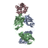
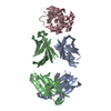
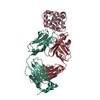
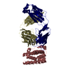
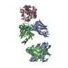
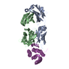
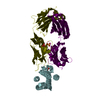
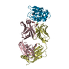
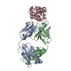
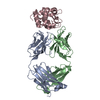
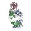
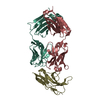
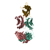
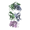
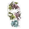
A: HyHEL10 heavy chain Fab fragment carrying I29F mutation. B: HyHEL10 light chain Fab fragment C: HyHEL10 heavy chain Fab fragment carrying I29F mutation. D: HyHEL10 light chain Fab fragment E: lysozyme isoform I (DEL-I) F: lysozyme isoform I (DEL-I) hetero molecules
Mass: 14428.297 Da / Num. of mol.: 2 / Source method: isolated from a natural source / Source: (natural) Anas platyrhynchos (mallard) / Tissue: egg white / References: UniProt: U3J0P1
-
Antibody , 2 types, 4 molecules ACBD
#1: Antibody
HyHEL10heavychainFabfragmentcarryingI29Fmutation.
Mass: 23171.660 Da / Num. of mol.: 2 / Fragment: DEL-I / Mutation: I29F Source method: isolated from a genetically manipulated source Source: (gene. exp.) Mus musculus (house mouse) / Cell line (production host): Expi293 / Production host: Homo sapiens (human) / References: exo-alpha-sialidase
#2: Antibody
HyHEL10lightchainFabfragment
Mass: 23206.500 Da / Num. of mol.: 2 Source method: isolated from a genetically manipulated source Source: (gene. exp.) Mus musculus (house mouse) / Gene: IgG light chain / Cell line (production host): Expi293 / Production host: Homo sapiens (human)
Resolution: 2.43→47.49 Å / Cor.coef. Fo:Fc: 0.916 / Cor.coef. Fo:Fc free: 0.895 / SU B: 28.682 / SU ML: 0.305 / SU R Cruickshank DPI: 0.5406 / Cross valid method: THROUGHOUT / σ(F): 0 / ESU R: 0.541 / ESU R Free: 0.314 Details: HYDROGENS HAVE BEEN ADDED IN THE RIDING POSITIONS U VALUES : WITH TLS ADDED
Rfactor
Num. reflection
% reflection
Selection details
Rfree
0.2959
2326
4.9 %
RANDOM
Rwork
0.2621
-
-
-
obs
0.2638
44719
99.78 %
-
Solvent computation
Ion probe radii: 0.8 Å / Shrinkage radii: 0.8 Å / VDW probe radii: 1.2 Å
In the structure databanks used in Yorodumi, some data are registered as the other names, "COVID-19 virus" and "2019-nCoV". Here are the details of the virus and the list of structure data.
Jan 31, 2019. EMDB accession codes are about to change! (news from PDBe EMDB page)
EMDB accession codes are about to change! (news from PDBe EMDB page)
The allocation of 4 digits for EMDB accession codes will soon come to an end. Whilst these codes will remain in use, new EMDB accession codes will include an additional digit and will expand incrementally as the available range of codes is exhausted. The current 4-digit format prefixed with “EMD-” (i.e. EMD-XXXX) will advance to a 5-digit format (i.e. EMD-XXXXX), and so on. It is currently estimated that the 4-digit codes will be depleted around Spring 2019, at which point the 5-digit format will come into force.
The EM Navigator/Yorodumi systems omit the EMD- prefix.
Related info.:Q: What is EMD? / ID/Accession-code notation in Yorodumi/EM Navigator
Yorodumi is a browser for structure data from EMDB, PDB, SASBDB, etc.
This page is also the successor to EM Navigator detail page, and also detail information page/front-end page for Omokage search.
The word "yorodu" (or yorozu) is an old Japanese word meaning "ten thousand". "mi" (miru) is to see.
Related info.:EMDB / PDB / SASBDB / Comparison of 3 databanks / Yorodumi Search / Aug 31, 2016. New EM Navigator & Yorodumi / Yorodumi Papers / Jmol/JSmol / Function and homology information / Changes in new EM Navigator and Yorodumi
 Movie
Movie Controller

 Yorodumi
 Open data
 Basic information
 Components
 Keywords
 Function and homology information

 X-RAY DIFFRACTION /
  Authors
 Australia, 1items
  Citation
 Structure visualization
 Downloads & links
 Other downloads
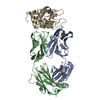


 PDBj
PDBj


 Assembly
 Homo sapiens (human) / References: exo-alpha-sialidase


 Mass: 35.453 Da / Num. of mol.: 4 / Source method: obtained synthetically / Formula: Cl
 Mass: 22.990 Da / Num. of mol.: 1 / Source method: obtained synthetically / Formula: Na
 Sample preparation
 Processing