 Movie
Movie Controller

[English] 日本語
 Yorodumi
- PDB-1mel: CRYSTAL STRUCTURE OF A CAMEL SINGLE-DOMAIN VH ANTIBODY FRAGMENT I... -
+ Open data

- Basic information
| Entry | Database: PDB / ID: 1mel | ||||||
|---|---|---|---|---|---|---|---|
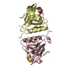
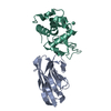
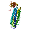
| Title | CRYSTAL STRUCTURE OF A CAMEL SINGLE-DOMAIN VH ANTIBODY FRAGMENT IN COMPLEX WITH LYSOZYME | ||||||
 Components Components |
| ||||||
 Keywords Keywords | COMPLEX (ANTIBODY/ANTIGEN) / CAMEL SINGLE-DOMAIN ANTI-LYSOZYME / COMPLEX (ANTIBODY-ANTIGEN) / COMPLEX (ANTIBODY-ANTIGEN) complex | ||||||
| Function / homology |  Function and homology information Function and homology informationLactose synthesis / Antimicrobial peptides / Neutrophil degranulation / beta-N-acetylglucosaminidase activity / cell wall macromolecule catabolic process / lysozyme / lysozyme activity / killing of cells of another organism / defense response to Gram-negative bacterium / defense response to bacterium ...Lactose synthesis / Antimicrobial peptides / Neutrophil degranulation / beta-N-acetylglucosaminidase activity / cell wall macromolecule catabolic process / lysozyme / lysozyme activity / killing of cells of another organism / defense response to Gram-negative bacterium / defense response to bacterium / defense response to Gram-positive bacterium / endoplasmic reticulum / : / identical protein binding / cytoplasm Similarity search - Function | ||||||
| Biological species |   | ||||||
| Method |  X-RAY DIFFRACTION / MOLECULAR REPLACEMENT / Resolution: 2.5 Å X-RAY DIFFRACTION / MOLECULAR REPLACEMENT / Resolution: 2.5 Å | ||||||
 Authors Authors | Desmyter, A. / Transue, T.R. / Arbabi Gharoudi, M. / Dao Thi, M. / Poortmans, F. / Hamers, R. / Muyldermans, S. / Wyns, L. | ||||||
 Citation Citation | Journal: Nat.Struct.Biol. / Year: 1996 Title: Crystal structure of a camel single-domain VH antibody fragment in complex with lysozyme. Authors: Desmyter, A. / Transue, T.R. / Ghahroudi, M.A. / Thi, M.H. / Poortmans, F. / Hamers, R. / Muyldermans, S. / Wyns, L. | ||||||
| History |
|
- Structure visualization
| Structure viewer | Molecule: MolmilJmol/JSmol |
|---|
- Downloads & links
-Download
| PDBx/mmCIF format | 1mel.cif.gz | 106.8 KB | Display | PDBx/mmCIF format |
|---|---|---|---|---|
| PDB format | pdb1mel.ent.gz | 82.9 KB | Display | PDB format |
| PDBx/mmJSON format | 1mel.json.gz | Tree view | PDBx/mmJSON format | |
| Others |  Other downloads Other downloads |
-Validation report
| Arichive directory | https://data.pdbj.org/pub/pdb/validation_reports/me/1melftp://data.pdbj.org/pub/pdb/validation_reports/me/1mel | HTTPS FTP |
|---|
-Related structure data


-Links
 PDBj
PDBj


- Assembly
| Deposited unit | 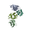
| ||||||||||||
|---|---|---|---|---|---|---|---|---|---|---|---|---|---|
| 1 | 
| ||||||||||||
| 2 | 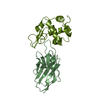
| ||||||||||||
| Unit cell |
| ||||||||||||
| Noncrystallographic symmetry (NCS) | NCS oper:
| ||||||||||||
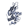
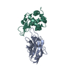
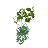
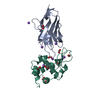
| Details | THE ASYMMETRIC UNIT CONTAINS TWO INDEPENDENTLY REFINED COMPLEXES: ANTIBODY A WITH LYSOZYME L AND ANTIBODY B WITH LYSOZYME M. SUBTLE DIFFERENCES BETWEEN THE TWO COMPLEXES HAVE BEEN OBSERVED, BUT HAVE NOT YET BEEN CAREFULLY ANALYZED. |
-Components
| #1: Antibody | Mass: 15657.090 Da / Num. of mol.: 2 Source method: isolated from a genetically manipulated source Source: (gene. exp.)  #2: Protein | Mass: 14331.160 Da / Num. of mol.: 2 Source method: isolated from a genetically manipulated source Source: (gene. exp.) #3: Water | ChemComp-HOH / |  Mass: 18.015 Da / Num. of mol.: 33 / Source method: isolated from a natural source / Formula: H2O Mass: 18.015 Da / Num. of mol.: 33 / Source method: isolated from a natural source / Formula: H2OHas protein modification | Y | |
|---|
-Experimental details
-Experiment
| Experiment | Method: X-RAY DIFFRACTION / Number of used crystals: 2 |
|---|
- Sample preparation
| Crystal | Density Matthews: 2.27 Å3/Da / Density % sol: 45 % | ||||||||||||||||||||
|---|---|---|---|---|---|---|---|---|---|---|---|---|---|---|---|---|---|---|---|---|---|
| Crystal grow | Method: vapor diffusion, hanging drop / pH: 6.4 Details: HANGING DROP, pH 6.4, vapor diffusion - hanging drop | ||||||||||||||||||||
| Crystal grow | *PLUS Method: vapor diffusion, hanging drop | ||||||||||||||||||||
| Components of the solutions | *PLUS
|
-Data collection
| Diffraction | Mean temperature: 298 K |
|---|---|
| Diffraction source | Source: ROTATING ANODE / Type: OTHER / Wavelength: 1.5418 |
| Detector | Type: ENRAF-NONIUS FAST / Detector: DIFFRACTOMETER / Date: May 2, 1996 / Details: COLLIMATOR |
| Radiation | Monochromator: GRAPHITE(002) / Monochromatic (M) / Laue (L): M / Scattering type: x-ray |
| Radiation wavelength | Wavelength: 1.5418 Å / Relative weight: 1 |
| Reflection | Resolution: 2.5→10 Å / Num. obs: 17829 / % possible obs: 93.4 % / Observed criterion σ(I): 0 / Redundancy: 5.4 % / Rmerge(I) obs: 0.114 / Rsym value: 0.114 / Net I/σ(I): 6.1 |
| Reflection shell | Resolution: 2.5→2.56 Å / Redundancy: 3.2 % / Rmerge(I) obs: 0.42 / Mean I/σ(I) obs: 1.8 / % possible all: 84.8 |
| Reflection shell | *PLUS % possible obs: 84.8 % |
- Processing
| Software |
| ||||||||||||||||||||||||||||||||||||||||||||||||||||||||||||
|---|---|---|---|---|---|---|---|---|---|---|---|---|---|---|---|---|---|---|---|---|---|---|---|---|---|---|---|---|---|---|---|---|---|---|---|---|---|---|---|---|---|---|---|---|---|---|---|---|---|---|---|---|---|---|---|---|---|---|---|---|---|
| Refinement | Method to determine structure: MOLECULAR REPLACEMENT Starting model: PDB ENTRIES 1LYS AND 1IGM Resolution: 2.5→10 Å / σ(F): 0 Details: RESIDUES ASP B 73 - LYS B 76 WERE LEFT IN THE MODEL TO CONSTRAIN RESIDUES 72 AND 77.
| ||||||||||||||||||||||||||||||||||||||||||||||||||||||||||||
| Displacement parameters | Biso mean: 16.7 Å2 | ||||||||||||||||||||||||||||||||||||||||||||||||||||||||||||
| Refinement step | Cycle: LAST / Resolution: 2.5→10 Å
| ||||||||||||||||||||||||||||||||||||||||||||||||||||||||||||
| Refine LS restraints |
| ||||||||||||||||||||||||||||||||||||||||||||||||||||||||||||
| Software | *PLUS Name: X-PLOR / Version: 3.1 / Classification: refinement | ||||||||||||||||||||||||||||||||||||||||||||||||||||||||||||
| Refine LS restraints | *PLUS
|
