 Movie
Movie Controller


+ Open data

- Basic information
| Entry | Database: PDB / ID: 1jto | ||||||
|---|---|---|---|---|---|---|---|
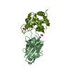
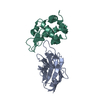
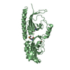
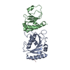
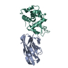
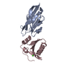
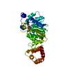
| Title | Degenerate interfaces in antigen-antibody complexes | ||||||
 Components Components |
| ||||||
 Keywords Keywords | Antibody / Hydrolase / immunoglobulin / heavy chain antibody / VHH / interface | ||||||
| Function / homology |  Function and homology information Function and homology informationLactose synthesis / Antimicrobial peptides / Neutrophil degranulation / beta-N-acetylglucosaminidase activity / cell wall macromolecule catabolic process / lysozyme / lysozyme activity / killing of cells of another organism / defense response to Gram-negative bacterium / defense response to bacterium ...Lactose synthesis / Antimicrobial peptides / Neutrophil degranulation / beta-N-acetylglucosaminidase activity / cell wall macromolecule catabolic process / lysozyme / lysozyme activity / killing of cells of another organism / defense response to Gram-negative bacterium / defense response to bacterium / defense response to Gram-positive bacterium / endoplasmic reticulum / : / identical protein binding / cytoplasm Similarity search - Function | ||||||
| Biological species |   | ||||||
| Method |  X-RAY DIFFRACTION / MOLECULAR REPLACEMENT / Resolution: 2.5 Å X-RAY DIFFRACTION / MOLECULAR REPLACEMENT / Resolution: 2.5 Å | ||||||
 Authors Authors | Decanniere, K. / Transue, T.R. / Desmyter, A. / Maes, D. / Muyldermans, S. / Wyns, L. | ||||||
 Citation Citation | Journal: J.Mol.Biol. / Year: 2001 Title: Degenerate interfaces in antigen-antibody complexes. Authors: Decanniere, K. / Transue, T.R. / Desmyter, A. / Maes, D. / Muyldermans, S. / Wyns, L. #1: Journal: Nat.Struct.Biol. / Year: 1996Title: Crystal structure of a camel single-domain VH antibody fragment in complex with lysozyme Authors: Desmyter, A. / Transue, T.R. / Ghahroudi, M.A. / Thi, M.H. / Poortmans, F. / Hamers, R. / Muyldermans, S. / Wyns, L. | ||||||
| History |
|
- Structure visualization
| Structure viewer | Molecule: MolmilJmol/JSmol |
|---|
- Downloads & links
-Download
| PDBx/mmCIF format | 1jto.cif.gz | 108.1 KB | Display | PDBx/mmCIF format |
|---|---|---|---|---|
| PDB format | pdb1jto.ent.gz | 84.1 KB | Display | PDB format |
| PDBx/mmJSON format | 1jto.json.gz | Tree view | PDBx/mmJSON format | |
| Others |  Other downloads Other downloads |
-Validation report
| Arichive directory | https://data.pdbj.org/pub/pdb/validation_reports/jt/1jtoftp://data.pdbj.org/pub/pdb/validation_reports/jt/1jto | HTTPS FTP |
|---|
-Related structure data
| Related structure data | 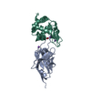 1jtpC 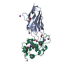 1jttC 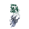 1melS S: Starting model for refinement C: citing same article ( |
|---|---|
| Similar structure data |


-Links
 PDBj
PDBj


- Assembly
| Deposited unit | 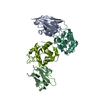
| ||||||||
|---|---|---|---|---|---|---|---|---|---|
| 1 | 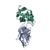
| ||||||||
| 2 | 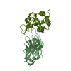
| ||||||||
| Unit cell |
|
-Components
| #1: Antibody | Mass: 15657.090 Da / Num. of mol.: 2 / Fragment: VH domain fragment Source method: isolated from a genetically manipulated source Source: (gene. exp.)  #2: Protein | Mass: 14331.160 Da / Num. of mol.: 2 / Fragment: Enzyme / Source method: isolated from a natural source / Details: Purchased from Sigma / Source: (natural) #3: Water | ChemComp-HOH / |  Mass: 18.015 Da / Num. of mol.: 49 / Source method: isolated from a natural source / Formula: H2O Mass: 18.015 Da / Num. of mol.: 49 / Source method: isolated from a natural source / Formula: H2OHas protein modification | Y | |
|---|
-Experimental details
-Experiment
| Experiment | Method: X-RAY DIFFRACTION / Number of used crystals: 2 |
|---|
- Sample preparation
| Crystal | Density Matthews: 2.27 Å3/Da / Density % sol: 45.77 % | |||||||||||||||
|---|---|---|---|---|---|---|---|---|---|---|---|---|---|---|---|---|
| Crystal grow | Temperature: 300 K / Method: vapor diffusion, hanging drop / pH: 6.4 Details: PEG 8000, potassium phosphate, , pH 6.4, VAPOR DIFFUSION, HANGING DROP, temperature 300K | |||||||||||||||
| Crystal grow | *PLUS pH: 5.6 / Method: vapor diffusion | |||||||||||||||
| Components of the solutions | *PLUS
|
-Data collection
| Diffraction | Mean temperature: 298 K |
|---|---|
| Diffraction source | Source: ROTATING ANODE / Type: ENRAF-NONIUS / Wavelength: 1.5418 Å |
| Detector | Type: ENRAF-NONIUS FAST / Detector: DIFFRACTOMETER / Date: May 1, 1996 |
| Radiation | Monochromator: Graphite single crystal / Protocol: SINGLE WAVELENGTH / Monochromatic (M) / Laue (L): M / Scattering type: x-ray |
| Radiation wavelength | Wavelength: 1.5418 Å / Relative weight: 1 |
| Reflection | Resolution: 2.5→20 Å / Num. all: 17829 / Num. obs: 17829 / % possible obs: 93.4 % / Observed criterion σ(F): 0 / Observed criterion σ(I): 0 / Redundancy: 5.4 % / Rmerge(I) obs: 0.114 / Rsym value: 0.114 / Net I/σ(I): 6.1 |
| Reflection shell | Resolution: 2.5→2.56 Å / Redundancy: 3.2 % / Rmerge(I) obs: 0.42 / Mean I/σ(I) obs: 1.8 / Num. unique all: 1170 / Rsym value: 0.42 / % possible all: 84.8 |
| Reflection | *PLUS |
- Processing
| Software |
| |||||||||||||||||||||
|---|---|---|---|---|---|---|---|---|---|---|---|---|---|---|---|---|---|---|---|---|---|---|
| Refinement | Method to determine structure: MOLECULAR REPLACEMENT Starting model: PDB ENTRY 1MEL Resolution: 2.5→20 Å / Isotropic thermal model: Overall anisotropic / Cross valid method: THROUGHOUT / σ(F): 0 / Stereochemistry target values: Engh & Huber
| |||||||||||||||||||||
| Displacement parameters |
| |||||||||||||||||||||
| Refinement step | Cycle: LAST / Resolution: 2.5→20 Å
| |||||||||||||||||||||
| Software | *PLUS Name: CNS / Classification: refinement | |||||||||||||||||||||
| Refinement | *PLUS Highest resolution: 2.5 Å / Lowest resolution: 20 Å / σ(F): 0 / Rfactor obs: 0.173 | |||||||||||||||||||||
| Solvent computation | *PLUS | |||||||||||||||||||||
| Displacement parameters | *PLUS | |||||||||||||||||||||
| Refine LS restraints | *PLUS
|
