 Movie
Movie Controller


+ Open data

- Basic information
| Entry | Database: PDB / ID: 1bvk | ||||||
|---|---|---|---|---|---|---|---|
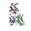
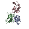
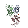
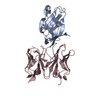
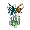
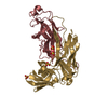
| Title | HUMANIZED ANTI-LYSOZYME FV COMPLEXED WITH LYSOZYME | ||||||
 Components Components |
| ||||||
 Keywords Keywords | COMPLEX (HUMANIZED ANTIBODY/HYDROLASE) / HUMANIZED ANTIBODY / ANTIBODY COMPLEX / FV / ANTI-LYSOZYME / COMPLEX (HUMANIZED ANTIBODY-HYDROLASE) / COMPLEX (HUMANIZED ANTIBODY-HYDROLASE) complex | ||||||
| Function / homology |  Function and homology information Function and homology informationLactose synthesis / Antimicrobial peptides / Neutrophil degranulation / beta-N-acetylglucosaminidase activity / cell wall macromolecule catabolic process / lysozyme / lysozyme activity / killing of cells of another organism / defense response to Gram-negative bacterium / defense response to bacterium ...Lactose synthesis / Antimicrobial peptides / Neutrophil degranulation / beta-N-acetylglucosaminidase activity / cell wall macromolecule catabolic process / lysozyme / lysozyme activity / killing of cells of another organism / defense response to Gram-negative bacterium / defense response to bacterium / defense response to Gram-positive bacterium / endoplasmic reticulum / : / identical protein binding / cytoplasm Similarity search - Function | ||||||
| Biological species |  Homo sapiens (human) Homo sapiens (human) | ||||||
| Method |  X-RAY DIFFRACTION / MOLECULAR REPLACEMENT / Resolution: 2.7 Å X-RAY DIFFRACTION / MOLECULAR REPLACEMENT / Resolution: 2.7 Å | ||||||
 Authors Authors | Holmes, M.A. / Buss, T.N. / Foote, J. | ||||||
 Citation Citation | Journal: J.Exp.Med. / Year: 1998 Title: Conformational correction mechanisms aiding antigen recognition by a humanized antibody. Authors: Holmes, M.A. / Buss, T.N. / Foote, J. #1: Journal: J.Immunol. / Year: 1997Title: Structural Consequences of Humanizing an Antibody Authors: Holmes, M.A. / Foote, J. #2: Journal: J.Mol.Biol. / Year: 1992Title: Antibody Framework Residues Affecting the Conformation of the Hypervariable Loops Authors: Foote, J. / Winter, G. | ||||||
| History |
|
- Structure visualization
| Structure viewer | Molecule: MolmilJmol/JSmol |
|---|
- Downloads & links
-Download
| PDBx/mmCIF format | 1bvk.cif.gz | 143.5 KB | Display | PDBx/mmCIF format |
|---|---|---|---|---|
| PDB format | pdb1bvk.ent.gz | 114.8 KB | Display | PDB format |
| PDBx/mmJSON format | 1bvk.json.gz | Tree view | PDBx/mmJSON format | |
| Others |  Other downloads Other downloads |
-Validation report
| Arichive directory | https://data.pdbj.org/pub/pdb/validation_reports/bv/1bvkftp://data.pdbj.org/pub/pdb/validation_reports/bv/1bvk | HTTPS FTP |
|---|
-Related structure data
| Related structure data | 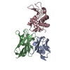 1vfbS S: Starting model for refinement |
|---|---|
| Similar structure data |


-Links
 PDBj
PDBj


- Assembly
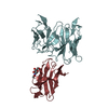
| Deposited unit | 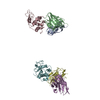
| ||||||||
|---|---|---|---|---|---|---|---|---|---|
| 1 | 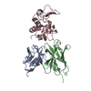
| ||||||||
| 2 | 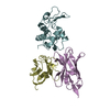
| ||||||||
| Unit cell |
| ||||||||
| Noncrystallographic symmetry (NCS) | NCS oper: (Code: given Matrix: (0.999987, 0.002639, 0.004298), Vector: |
-Components
| #1: Antibody | Mass: 11962.320 Da / Num. of mol.: 2 / Fragment: FV Source method: isolated from a genetically manipulated source Source: (gene. exp.) Homo sapiens (human) / Production host:  #2: Antibody | Mass: 12872.358 Da / Num. of mol.: 2 / Fragment: FV Source method: isolated from a genetically manipulated source Source: (gene. exp.) Homo sapiens (human) / Production host: #3: Protein | Mass: 14331.160 Da / Num. of mol.: 2 Source method: isolated from a genetically manipulated source Source: (gene. exp.) Has protein modification | Y | |
|---|
-Experimental details
-Experiment
| Experiment | Method: X-RAY DIFFRACTION / Number of used crystals: 1 |
|---|
- Sample preparation
| Crystal | Density Matthews: 2.67 Å3/Da / Density % sol: 53.9 % | ||||||||||||||||||||
|---|---|---|---|---|---|---|---|---|---|---|---|---|---|---|---|---|---|---|---|---|---|
| Crystal grow | pH: 6.5 / Details: pH 6.5 | ||||||||||||||||||||
| Crystal grow | *PLUS Method: vapor diffusion | ||||||||||||||||||||
| Components of the solutions | *PLUS
|
-Data collection
| Diffraction | Mean temperature: 277 K |
|---|---|
| Diffraction source | Source: ROTATING ANODE / Type: RIGAKU RUH2R / Wavelength: 1.5418 |
| Detector | Type: RIGAKU / Detector: IMAGE PLATE / Date: Nov 1, 1996 / Details: R-AXIS IIC |
| Radiation | Monochromator: GRAPHITE(002) / Monochromatic (M) / Laue (L): M / Scattering type: x-ray |
| Radiation wavelength | Wavelength: 1.5418 Å / Relative weight: 1 |
| Reflection | Resolution: 2.7→50 Å / Num. obs: 22379 / % possible obs: 93.2 % / Observed criterion σ(I): 0 / Redundancy: 5.4 % / Biso Wilson estimate: 44.1 Å2 / Rmerge(I) obs: 0.075 / Rsym value: 0.075 / Net I/σ(I): 9.6 |
| Reflection shell | Resolution: 2.7→2.75 Å / Rmerge(I) obs: 0.452 / Rsym value: 0.452 / % possible all: 73.5 |
| Reflection | *PLUS Num. measured all: 120820 |
| Reflection shell | *PLUS % possible obs: 73.5 % |
- Processing
| Software |
| ||||||||||||||||||||||||||||||||||||||||||||||||||||||||||||||||||||||||||||||||
|---|---|---|---|---|---|---|---|---|---|---|---|---|---|---|---|---|---|---|---|---|---|---|---|---|---|---|---|---|---|---|---|---|---|---|---|---|---|---|---|---|---|---|---|---|---|---|---|---|---|---|---|---|---|---|---|---|---|---|---|---|---|---|---|---|---|---|---|---|---|---|---|---|---|---|---|---|---|---|---|---|---|
| Refinement | Method to determine structure: MOLECULAR REPLACEMENT Starting model: HULYS STRUCTURE, AND LYSOZYME FROM PDB ENTRY 1VFB Resolution: 2.7→10 Å / Data cutoff high absF: 100000 / Data cutoff low absF: 0.1 / Isotropic thermal model: RESTRAINED / Cross valid method: THROUGHOUT / σ(F): 2 Details: A FEW CYCLES OF TNT REFINEMENT WERE INTERSPERSED WITH THE X-PLOR CYCLES.
| ||||||||||||||||||||||||||||||||||||||||||||||||||||||||||||||||||||||||||||||||
| Displacement parameters | Biso mean: 31.1 Å2 | ||||||||||||||||||||||||||||||||||||||||||||||||||||||||||||||||||||||||||||||||
| Refine analyze |
| ||||||||||||||||||||||||||||||||||||||||||||||||||||||||||||||||||||||||||||||||
| Refinement step | Cycle: LAST / Resolution: 2.7→10 Å
| ||||||||||||||||||||||||||||||||||||||||||||||||||||||||||||||||||||||||||||||||
| Refine LS restraints |
| ||||||||||||||||||||||||||||||||||||||||||||||||||||||||||||||||||||||||||||||||
| LS refinement shell | Resolution: 2.7→2.82 Å / Total num. of bins used: 8
| ||||||||||||||||||||||||||||||||||||||||||||||||||||||||||||||||||||||||||||||||
| Xplor file |
| ||||||||||||||||||||||||||||||||||||||||||||||||||||||||||||||||||||||||||||||||
| Software | *PLUS Name: X-PLOR / Version: 3.8 / Classification: refinement | ||||||||||||||||||||||||||||||||||||||||||||||||||||||||||||||||||||||||||||||||
| Refinement | *PLUS | ||||||||||||||||||||||||||||||||||||||||||||||||||||||||||||||||||||||||||||||||
| Solvent computation | *PLUS | ||||||||||||||||||||||||||||||||||||||||||||||||||||||||||||||||||||||||||||||||
| Displacement parameters | *PLUS | ||||||||||||||||||||||||||||||||||||||||||||||||||||||||||||||||||||||||||||||||
| Refine LS restraints | *PLUS
| ||||||||||||||||||||||||||||||||||||||||||||||||||||||||||||||||||||||||||||||||
| LS refinement shell | *PLUS Rfactor obs: 0.326 |
