 Movie
Movie Controller


+ Open data

- Basic information
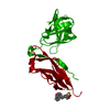
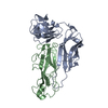
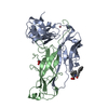
| Entry | Database: PDB / ID: 2uy7 | ||||||
|---|---|---|---|---|---|---|---|
| Title | Crystal structure of the P pilus rod subunit PapA | ||||||
 Components Components |
| ||||||
 Keywords Keywords | CHAPERONE / DONOR STRAND COMPLEMENTATION / PILI/N-TERMINAL EXTENSION / PILUS BIOGENESIS / DONOR-STRAND EXCHANGE / NTE / DSC / DSE / PAPA / PAPD / FIMBRIA | ||||||
| Function / homology |  Function and homology information Function and homology informationcell adhesion involved in single-species biofilm formation / pilus / cell wall organization / outer membrane-bounded periplasmic space / extracellular region Similarity search - Function | ||||||
| Biological species |  | ||||||
| Method |  X-RAY DIFFRACTION / SYNCHROTRON / MOLECULAR REPLACEMENT / Resolution: 2.6 Å X-RAY DIFFRACTION / SYNCHROTRON / MOLECULAR REPLACEMENT / Resolution: 2.6 Å | ||||||
 Authors Authors | Verger, D. / Bullitt, E. / Hultgren, S.J. / Waksman, G. | ||||||
 Citation Citation | Journal: Plos Pathog. / Year: 2007 Title: Crystal Structure of the P Pilus Rod Subunit Papa. Authors: Verger, D. / Bullitt, E. / Hultgren, S.J. / Waksman, G. | ||||||
| History |
| ||||||
| Remark 700 | SHEET THE SHEET STRUCTURE OF THIS MOLECULE IS BIFURCATED. IN ORDER TO REPRESENT THIS FEATURE IN ... SHEET THE SHEET STRUCTURE OF THIS MOLECULE IS BIFURCATED. IN ORDER TO REPRESENT THIS FEATURE IN THE SHEET RECORDS BELOW, TWO SHEETS ARE DEFINED. |
- Structure visualization
| Structure viewer | Molecule: MolmilJmol/JSmol |
|---|
- Downloads & links
-Download
| PDBx/mmCIF format | 2uy7.cif.gz | 286.7 KB | Display | PDBx/mmCIF format |
|---|---|---|---|---|
| PDB format | pdb2uy7.ent.gz | 231.9 KB | Display | PDB format |
| PDBx/mmJSON format | 2uy7.json.gz | Tree view | PDBx/mmJSON format | |
| Others |  Other downloads Other downloads |
-Validation report
| Arichive directory | https://data.pdbj.org/pub/pdb/validation_reports/uy/2uy7ftp://data.pdbj.org/pub/pdb/validation_reports/uy/2uy7 | HTTPS FTP |
|---|
-Related structure data
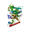
| Related structure data |  2uy6C  1pdkS C: citing same article ( S: Starting model for refinement |
|---|---|
| Similar structure data |


-Links
 PDBj
PDBj
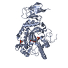
- Assembly
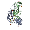
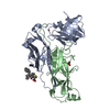
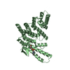
| Deposited unit | 
| ||||||||
|---|---|---|---|---|---|---|---|---|---|
| 1 | 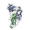
| ||||||||
| 2 | 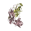
| ||||||||
| 3 | 
| ||||||||
| 4 | 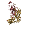
| ||||||||
| Unit cell |
|
-Components
| #1: Protein | Mass: 24589.895 Da / Num. of mol.: 4 Source method: isolated from a genetically manipulated source Details: WILD TYPE P PILUS CHAPERONE PAPD / Source: (gene. exp.) #2: Protein | Mass: 16625.396 Da / Num. of mol.: 4 / Mutation: YES Source method: isolated from a genetically manipulated source Details: P PILUS ROD SUBUNIT PAPA / Source: (gene. exp.) #3: Chemical | ChemComp-SO4 /   Mass: 96.063 Da / Num. of mol.: 13 / Source method: obtained synthetically / Formula: SO4 Mass: 96.063 Da / Num. of mol.: 13 / Source method: obtained synthetically / Formula: SO4#4: Water | ChemComp-HOH / |  Mass: 18.015 Da / Num. of mol.: 232 / Source method: isolated from a natural source / Formula: H2O Mass: 18.015 Da / Num. of mol.: 232 / Source method: isolated from a natural source / Formula: H2OCompound details | ENGINEERED RESIDUE IN CHAIN B, GLY 37 TO ASN ENGINEERED RESIDUE IN CHAIN D, GLY 37 TO ASN ...ENGINEERED | Has protein modification | Y | Sequence details | MATURE PROTEIN SEQUENCE AFTER CLIVAGE OF SIGNAL SEQUENCE | |
|---|
-Experimental details
-Experiment
| Experiment | Method: X-RAY DIFFRACTION / Number of used crystals: 1 |
|---|
- Sample preparation
| Crystal | Density Matthews: 4.24 Å3/Da / Density % sol: 71 % |
|---|---|
| Crystal grow | pH: 5.6 / Details: 2M AMMONIUM SULFATE, 0.1M SODIUM ACETATE PH = 5.6 |
-Data collection
| Diffraction | Mean temperature: 100 K |
|---|---|
| Diffraction source | Source: SYNCHROTRON / Site: ESRF  / Beamline: ID29 / Wavelength: 0.976 / Beamline: ID29 / Wavelength: 0.976 |
| Detector | Type: ADSC CCD / Detector: CCD / Date: Feb 25, 2005 |
| Radiation | Protocol: SINGLE WAVELENGTH / Monochromatic (M) / Laue (L): M / Scattering type: x-ray |
| Radiation wavelength | Wavelength: 0.976 Å / Relative weight: 1 |
| Reflection | Resolution: 2.6→75.8 Å / Num. obs: 88336 / % possible obs: 99.8 % / Observed criterion σ(I): 0 / Redundancy: 5.2 % / Biso Wilson estimate: 53.5 Å2 / Rmerge(I) obs: 0.09 / Net I/σ(I): 7.3 |
| Reflection shell | Resolution: 2.6→2.74 Å / Redundancy: 5 % / Rmerge(I) obs: 0.45 / Mean I/σ(I) obs: 1.7 / % possible all: 99.7 |
- Processing
| Software |
| ||||||||||||||||||||||||||||||||||||||||||||||||||||||||||||
|---|---|---|---|---|---|---|---|---|---|---|---|---|---|---|---|---|---|---|---|---|---|---|---|---|---|---|---|---|---|---|---|---|---|---|---|---|---|---|---|---|---|---|---|---|---|---|---|---|---|---|---|---|---|---|---|---|---|---|---|---|---|
| Refinement | Method to determine structure: MOLECULAR REPLACEMENT Starting model: PDB ENTRY 1PDK Resolution: 2.6→75.8 Å / Data cutoff high absF: 10000 / Isotropic thermal model: RESTRAINED / Cross valid method: THROUGHOUT / σ(F): 0 / Stereochemistry target values: MAXIMUM LIKELIHOOD Details: THE 4 COMPLEXES IN THE ASYMETRIC UNIT WERE RETRAINED BY NCS, USING THE MOST SIMILAR PARTS OF THE CORE STRUCTURE (MAIN CHAIN)
| ||||||||||||||||||||||||||||||||||||||||||||||||||||||||||||
| Solvent computation | Bsol: 38.4771 Å2 / ksol: 0.36868 e/Å3 | ||||||||||||||||||||||||||||||||||||||||||||||||||||||||||||
| Refinement step | Cycle: LAST / Resolution: 2.6→75.8 Å
| ||||||||||||||||||||||||||||||||||||||||||||||||||||||||||||
| Refine LS restraints |
| ||||||||||||||||||||||||||||||||||||||||||||||||||||||||||||
| LS refinement shell | Resolution: 2.6→2.62 Å / Total num. of bins used: 50
| ||||||||||||||||||||||||||||||||||||||||||||||||||||||||||||
| Xplor file |
|
