 Movie
Movie Controller

[English] 日本語
 Yorodumi
- PDB-1mhp: Crystal structure of a chimeric alpha1 integrin I-domain in compl... -
+ Open data

- Basic information
| Entry | Database: PDB / ID: 1mhp | ||||||
|---|---|---|---|---|---|---|---|
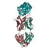
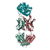
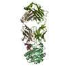
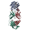
| Title | Crystal structure of a chimeric alpha1 integrin I-domain in complex with the Fab fragment of a humanized neutralizing antibody | ||||||
 Components Components |
| ||||||
 Keywords Keywords | IMMUNE SYSTEM / integrin / cell adhesion / receptor / antibody | ||||||
| Function / homology |  Function and homology information Function and homology informationIntegrin cell surface interactions / Smooth Muscle Contraction / integrin alpha1-beta1 complex / cellular extravasation / collagen binding involved in cell-matrix adhesion / phosphatase activator activity / basal part of cell / negative regulation of epidermal growth factor receptor signaling pathway / collagen binding / neuron projection morphogenesis ...Integrin cell surface interactions / Smooth Muscle Contraction / integrin alpha1-beta1 complex / cellular extravasation / collagen binding involved in cell-matrix adhesion / phosphatase activator activity / basal part of cell / negative regulation of epidermal growth factor receptor signaling pathway / collagen binding / neuron projection morphogenesis / neutrophil chemotaxis / acrosomal vesicle / integrin-mediated signaling pathway / cell-matrix adhesion / cell chemotaxis / cell-cell adhesion / vasodilation / positive regulation of neuron apoptotic process / signaling receptor activity / protein phosphatase binding / perikaryon / positive regulation of MAPK cascade / cell adhesion / negative regulation of cell population proliferation / external side of plasma membrane / signaling receptor binding / focal adhesion / cell surface / metal ion binding Similarity search - Function | ||||||
| Biological species |  | ||||||
| Method |  X-RAY DIFFRACTION / SYNCHROTRON / MOLECULAR REPLACEMENT / Resolution: 2.8 Å X-RAY DIFFRACTION / SYNCHROTRON / MOLECULAR REPLACEMENT / Resolution: 2.8 Å | ||||||
 Authors Authors | Karpusas, M. / Taylor, F. / Ferrant, J. / Weinreb, P. / Garber, E. | ||||||
 Citation Citation | Journal: J.Mol.Biol. / Year: 2003 Title: Crystal Structure of the alpha 1 beta 1 Integrin I Domain in Complex with an Antibody Fab Fragment Authors: Karpusas, M. / Ferrant, J. / Weinreb, P. / Carmillo, A. / Taylor, F. / Garber, E. | ||||||
| History |
| ||||||
| Remark 999 | SEQUENCE AN APPROPRIATE SEQUENCE DATABASE REFERENCE WAS NOT AVAILABLE AT THE TIME OF PROCESSING FOR ...SEQUENCE AN APPROPRIATE SEQUENCE DATABASE REFERENCE WAS NOT AVAILABLE AT THE TIME OF PROCESSING FOR CHAINS H AND X (Fab fragment, heavy chain) AND FOR CHAINS L AND Y (FAB FRAGMENT, light chain). |
- Structure visualization
| Structure viewer | Molecule: MolmilJmol/JSmol |
|---|
- Downloads & links
-Download
| PDBx/mmCIF format | 1mhp.cif.gz | 202.3 KB | Display | PDBx/mmCIF format |
|---|---|---|---|---|
| PDB format | pdb1mhp.ent.gz | 161.2 KB | Display | PDB format |
| PDBx/mmJSON format | 1mhp.json.gz | Tree view | PDBx/mmJSON format | |
| Others |  Other downloads Other downloads |
-Validation report
| Arichive directory | https://data.pdbj.org/pub/pdb/validation_reports/mh/1mhpftp://data.pdbj.org/pub/pdb/validation_reports/mh/1mhp | HTTPS FTP |
|---|
-Related structure data
| Similar structure data |
|---|


-Links
 PDBj
PDBj


- Assembly
| Deposited unit | 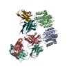
| ||||||||
|---|---|---|---|---|---|---|---|---|---|
| 1 | 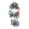
| ||||||||
| 2 | 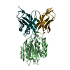
| ||||||||
| Unit cell |
|
-Components
| #1: Protein | Mass: 21629.518 Da / Num. of mol.: 2 / Fragment: alpha1 I-domain / Mutation: G217V, R218Q, Q219R, L222R Source method: isolated from a genetically manipulated source Source: (gene. exp.)  #2: Antibody | Mass: 23141.895 Da / Num. of mol.: 2 Source method: isolated from a genetically manipulated source Source: (gene. exp.)  Cricetulus griseus (Chinese hamster) Cricetulus griseus (Chinese hamster)#3: Antibody | Mass: 23201.668 Da / Num. of mol.: 2 Source method: isolated from a genetically manipulated source Source: (gene. exp.) Cricetulus griseus (Chinese hamster)#4: Chemical |   Mass: 54.938 Da / Num. of mol.: 2 / Source method: obtained synthetically / Formula: Mn Mass: 54.938 Da / Num. of mol.: 2 / Source method: obtained synthetically / Formula: MnHas protein modification | Y | |
|---|
-Experimental details
-Experiment
| Experiment | Method: X-RAY DIFFRACTION / Number of used crystals: 1 |
|---|
- Sample preparation
| Crystal | Density Matthews: 2.67 Å3/Da / Density % sol: 53.91 % | |||||||||||||||||||||||||||||||||||||||||||||||||
|---|---|---|---|---|---|---|---|---|---|---|---|---|---|---|---|---|---|---|---|---|---|---|---|---|---|---|---|---|---|---|---|---|---|---|---|---|---|---|---|---|---|---|---|---|---|---|---|---|---|---|
| Crystal grow | Temperature: 293 K / Method: vapor diffusion, hanging drop / pH: 7.4 Details: PEG 1500, Tris, NaCl, beta-mercaptoethanol, pH 7.4, VAPOR DIFFUSION, HANGING DROP, temperature 293.0K | |||||||||||||||||||||||||||||||||||||||||||||||||
| Crystal grow | *PLUS Temperature: 20 ℃ / Method: vapor diffusion | |||||||||||||||||||||||||||||||||||||||||||||||||
| Components of the solutions | *PLUS
|
-Data collection
| Diffraction | Mean temperature: 100 K |
|---|---|
| Diffraction source | Source: SYNCHROTRON / Site: NSLS  / Beamline: X4A / Wavelength: 0.979 Å / Beamline: X4A / Wavelength: 0.979 Å |
| Detector | Type: ADSC QUANTUM 4 / Detector: CCD / Date: Oct 5, 2001 |
| Radiation | Protocol: SINGLE WAVELENGTH / Monochromatic (M) / Laue (L): M / Scattering type: x-ray |
| Radiation wavelength | Wavelength: 0.979 Å / Relative weight: 1 |
| Reflection | Resolution: 2.8→35 Å / Num. all: 36501 / Num. obs: 35275 / % possible obs: 96.6 % / Observed criterion σ(F): 1 / Observed criterion σ(I): 1 / Rmerge(I) obs: 0.083 / Net I/σ(I): 11.92 |
| Reflection shell | Resolution: 2.8→2.9 Å / Rmerge(I) obs: 0.309 / Mean I/σ(I) obs: 2.29 / % possible all: 87.7 |
| Reflection | *PLUS Lowest resolution: 500 Å |
| Reflection shell | *PLUS % possible obs: 87.7 % |
- Processing
| Software |
| ||||||||||||||||||||
|---|---|---|---|---|---|---|---|---|---|---|---|---|---|---|---|---|---|---|---|---|---|
| Refinement | Method to determine structure: MOLECULAR REPLACEMENT / Resolution: 2.8→35 Å / σ(F): 2 / Stereochemistry target values: Engh & Huber
| ||||||||||||||||||||
| Refinement step | Cycle: LAST / Resolution: 2.8→35 Å
| ||||||||||||||||||||
| Refine LS restraints |
| ||||||||||||||||||||
| Refinement | *PLUS Lowest resolution: 500 Å | ||||||||||||||||||||
| Solvent computation | *PLUS | ||||||||||||||||||||
| Displacement parameters | *PLUS | ||||||||||||||||||||
| Refine LS restraints | *PLUS
|
