 Movie
Movie Controller

[English] 日本語
 Yorodumi
- PDB-3qxm: Crystal Structure of Human GluK2 Ligand-Binding Core in Complex w... -
+ Open data

- Basic information
| Entry | Database: PDB / ID: 3qxm | ||||||
|---|---|---|---|---|---|---|---|
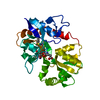
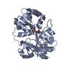
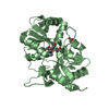
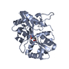
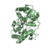
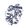
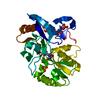
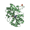
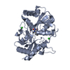
| Title | Crystal Structure of Human GluK2 Ligand-Binding Core in Complex with Novel Marine-Derived Toxins, Neodysiherbaine A | ||||||
 Components Components | Glutamate receptor ionotropic, kainate 2 | ||||||
 Keywords Keywords | MEMBRANE PROTEIN / two domains / ligand-binding | ||||||
| Function / homology |  Function and homology information Function and homology informationActivation of Na-permeable kainate receptors / mossy fiber rosette / detection of cold stimulus involved in thermoception / kainate selective glutamate receptor complex / regulation of short-term neuronal synaptic plasticity / ubiquitin conjugating enzyme binding / negative regulation of synaptic transmission, glutamatergic / regulation of JNK cascade / Activation of Ca-permeable Kainate Receptor / glutamate receptor signaling pathway ...Activation of Na-permeable kainate receptors / mossy fiber rosette / detection of cold stimulus involved in thermoception / kainate selective glutamate receptor complex / regulation of short-term neuronal synaptic plasticity / ubiquitin conjugating enzyme binding / negative regulation of synaptic transmission, glutamatergic / regulation of JNK cascade / Activation of Ca-permeable Kainate Receptor / glutamate receptor signaling pathway / receptor clustering / kainate selective glutamate receptor activity / extracellularly glutamate-gated ion channel activity / modulation of excitatory postsynaptic potential / positive regulation of synaptic transmission / glutamate-gated receptor activity / glutamate-gated calcium ion channel activity / dendrite cytoplasm / ligand-gated monoatomic ion channel activity involved in regulation of presynaptic membrane potential / hippocampal mossy fiber to CA3 synapse / SNARE binding / PDZ domain binding / synaptic transmission, glutamatergic / transmitter-gated monoatomic ion channel activity involved in regulation of postsynaptic membrane potential / postsynaptic density membrane / modulation of chemical synaptic transmission / terminal bouton / positive regulation of neuron apoptotic process / neuron apoptotic process / presynaptic membrane / scaffold protein binding / chemical synaptic transmission / perikaryon / ubiquitin protein ligase binding / glutamatergic synapse / identical protein binding / plasma membrane Similarity search - Function | ||||||
| Biological species |  Homo sapiens (human) Homo sapiens (human) | ||||||
| Method |  X-RAY DIFFRACTION / SYNCHROTRON / MOLECULAR REPLACEMENT / Resolution: 1.65 Å X-RAY DIFFRACTION / SYNCHROTRON / MOLECULAR REPLACEMENT / Resolution: 1.65 Å | ||||||
 Authors Authors | Unno, M. / Sasaki, M. / Ikeda-Saito, M. | ||||||
 Citation Citation | Journal: J.Mol.Biol. / Year: 2011 Title: Binding and Selectivity of the Marine Toxin Neodysiherbaine A and Its Synthetic Analogues to GluK1 and GluK2 Kainate Receptors. Authors: Unno, M. / Shinohara, M. / Takayama, K. / Tanaka, H. / Teruya, K. / Doh-Ura, K. / Sakai, R. / Sasaki, M. / Ikeda-Saito, M. | ||||||
| History |
|
- Structure visualization
| Structure viewer | Molecule: MolmilJmol/JSmol |
|---|
- Downloads & links
-Download
| PDBx/mmCIF format | 3qxm.cif.gz | 221.6 KB | Display | PDBx/mmCIF format |
|---|---|---|---|---|
| PDB format | pdb3qxm.ent.gz | 178.3 KB | Display | PDB format |
| PDBx/mmJSON format | 3qxm.json.gz | Tree view | PDBx/mmJSON format | |
| Others |  Other downloads Other downloads |
-Validation report
| Arichive directory | https://data.pdbj.org/pub/pdb/validation_reports/qx/3qxmftp://data.pdbj.org/pub/pdb/validation_reports/qx/3qxm | HTTPS FTP |
|---|
-Related structure data
| Related structure data | 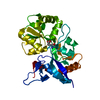 2znsC  2zntC 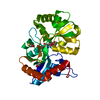 2znuC  3fuzC  3fv1C  3fv2C  3fvgC  3fvkC  3fvnC 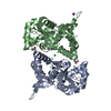 3g3fS C: citing same article ( S: Starting model for refinement |
|---|---|
| Similar structure data |


-Links
 PDBj
PDBj


- Assembly
| Deposited unit | 
| ||||||||
|---|---|---|---|---|---|---|---|---|---|
| 1 | 
| ||||||||
| 2 | 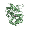
| ||||||||
| Unit cell |
|
-Components
| #1: Protein | Mass: 29286.527 Da / Num. of mol.: 2 / Fragment: UNP RESIDUES 429-544,UNP RESIDUES 667-806 Source method: isolated from a genetically manipulated source Details: The fusion protein of RESIDUES 429-544 of GluR-6, the linker GLY-THR and 667-806 of GluR-6 Source: (gene. exp.) Homo sapiens (human) / Gene: GRIK2, GLUR6 / Plasmid: pET28a / Production host:  #2: Chemical |   Mass: 291.255 Da / Num. of mol.: 2 / Source method: obtained synthetically / Formula: C11H17NO8 Mass: 291.255 Da / Num. of mol.: 2 / Source method: obtained synthetically / Formula: C11H17NO8#3: Water | ChemComp-HOH / |  Mass: 18.015 Da / Num. of mol.: 474 / Source method: isolated from a natural source / Formula: H2O Mass: 18.015 Da / Num. of mol.: 474 / Source method: isolated from a natural source / Formula: H2OHas protein modification | Y | |
|---|
-Experimental details
-Experiment
| Experiment | Method: X-RAY DIFFRACTION / Number of used crystals: 1 |
|---|
- Sample preparation
| Crystal | Density Matthews: 2.37 Å3/Da / Density % sol: 48.1 % |
|---|---|
| Crystal grow | Temperature: 303 K / Method: vapor diffusion, hanging drop / pH: 6.5 Details: 18-22% PEG4000, 2mM Tris, 1mM EDTA, 10mM NaCl, 10mM Sodium L-ascorbate monohydrate, pH 6.5, VAPOR DIFFUSION, HANGING DROP, temperature 303K |
-Data collection
| Diffraction | Mean temperature: 95 K |
|---|---|
| Diffraction source | Source: SYNCHROTRON / Site: Photon Factory  / Beamline: AR-NE3A / Wavelength: 1 Å / Beamline: AR-NE3A / Wavelength: 1 Å |
| Detector | Type: ADSC QUANTUM 270 / Detector: CCD / Date: Nov 9, 2009 |
| Radiation | Monochromator: Si111 / Protocol: SINGLE WAVELENGTH / Monochromatic (M) / Laue (L): M / Scattering type: x-ray |
| Radiation wavelength | Wavelength: 1 Å / Relative weight: 1 |
| Reflection | Resolution: 1.65→50 Å / Num. all: 67908 / Num. obs: 62566 / % possible obs: 96.11 % / Observed criterion σ(F): 0 / Observed criterion σ(I): 0 |
| Reflection shell | Resolution: 1.65→1.68 Å / % possible all: 75.3 |
- Processing
| Software |
| |||||||||||||||||||||||||||||||||||||||||||||||||||||||||||||||||||||||||||||||||||||||||||||||||||||||||||||||||||||||||||||||||||||||||||||||||||||||||||||||||||||||||||||||
|---|---|---|---|---|---|---|---|---|---|---|---|---|---|---|---|---|---|---|---|---|---|---|---|---|---|---|---|---|---|---|---|---|---|---|---|---|---|---|---|---|---|---|---|---|---|---|---|---|---|---|---|---|---|---|---|---|---|---|---|---|---|---|---|---|---|---|---|---|---|---|---|---|---|---|---|---|---|---|---|---|---|---|---|---|---|---|---|---|---|---|---|---|---|---|---|---|---|---|---|---|---|---|---|---|---|---|---|---|---|---|---|---|---|---|---|---|---|---|---|---|---|---|---|---|---|---|---|---|---|---|---|---|---|---|---|---|---|---|---|---|---|---|---|---|---|---|---|---|---|---|---|---|---|---|---|---|---|---|---|---|---|---|---|---|---|---|---|---|---|---|---|---|---|---|---|---|
| Refinement | Method to determine structure: MOLECULAR REPLACEMENT Starting model: 3G3F Resolution: 1.65→29.92 Å / Cor.coef. Fo:Fc: 0.966 / Cor.coef. Fo:Fc free: 0.95 / SU B: 4.619 / SU ML: 0.073 / Cross valid method: THROUGHOUT / ESU R Free: 0.105 / Stereochemistry target values: MAXIMUM LIKELIHOOD / Details: HYDROGENS HAVE BEEN ADDED IN THE RIDING POSITIONS
| |||||||||||||||||||||||||||||||||||||||||||||||||||||||||||||||||||||||||||||||||||||||||||||||||||||||||||||||||||||||||||||||||||||||||||||||||||||||||||||||||||||||||||||||
| Solvent computation | Ion probe radii: 0.8 Å / Shrinkage radii: 0.8 Å / VDW probe radii: 1.4 Å / Solvent model: MASK | |||||||||||||||||||||||||||||||||||||||||||||||||||||||||||||||||||||||||||||||||||||||||||||||||||||||||||||||||||||||||||||||||||||||||||||||||||||||||||||||||||||||||||||||
| Displacement parameters | Biso mean: 27.369 Å2
| |||||||||||||||||||||||||||||||||||||||||||||||||||||||||||||||||||||||||||||||||||||||||||||||||||||||||||||||||||||||||||||||||||||||||||||||||||||||||||||||||||||||||||||||
| Refinement step | Cycle: LAST / Resolution: 1.65→29.92 Å
| |||||||||||||||||||||||||||||||||||||||||||||||||||||||||||||||||||||||||||||||||||||||||||||||||||||||||||||||||||||||||||||||||||||||||||||||||||||||||||||||||||||||||||||||
| Refine LS restraints |
| |||||||||||||||||||||||||||||||||||||||||||||||||||||||||||||||||||||||||||||||||||||||||||||||||||||||||||||||||||||||||||||||||||||||||||||||||||||||||||||||||||||||||||||||
| LS refinement shell | Resolution: 1.65→1.693 Å / Total num. of bins used: 20
| |||||||||||||||||||||||||||||||||||||||||||||||||||||||||||||||||||||||||||||||||||||||||||||||||||||||||||||||||||||||||||||||||||||||||||||||||||||||||||||||||||||||||||||||
| Refinement TLS params. | Method: refined / Refine-ID: X-RAY DIFFRACTION
| |||||||||||||||||||||||||||||||||||||||||||||||||||||||||||||||||||||||||||||||||||||||||||||||||||||||||||||||||||||||||||||||||||||||||||||||||||||||||||||||||||||||||||||||
| Refinement TLS group |
|
