[English] 日本語
Yorodumi
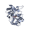
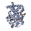
- PDB-6f28: Crystal structure of the kainate receptor GluK3 ligand binding do... -

+
Open data


ID or keywords:

Loading...

-
Basic information

Entry
Database: PDB / ID: 6f28
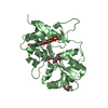
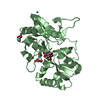
Title
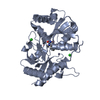
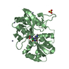
Crystal structure of the kainate receptor GluK3 ligand binding domain in complex with (S)-1-[2'-Amino-2'-carboxyethyl]-6-methyl-5,7-dihydropyrrolo[3,4-d]pyrimidin-2,4(1H,3H)-dione at resolution 2.4A
ComponentsGlutamate receptor ionotropic, kainate 3,Glutamate receptor ionotropic, kainate 3
KeywordsMEMBRANE PROTEIN / ionotropic glutamate receptor / GluK3 / ligand binding domain / membrane protein.
Function / homology
Function and homology information


Presynaptic function of Kainate receptors / adenylate cyclase inhibiting G protein-coupled glutamate receptor activity / G protein-coupled glutamate receptor signaling pathway / kainate selective glutamate receptor complex / Activation of Ca-permeable Kainate Receptor / negative regulation of synaptic transmission, glutamatergic / glutamate receptor signaling pathway / glutamate receptor activity / kainate selective glutamate receptor activity / glutamate-gated receptor activity ...Presynaptic function of Kainate receptors / adenylate cyclase inhibiting G protein-coupled glutamate receptor activity / G protein-coupled glutamate receptor signaling pathway / kainate selective glutamate receptor complex / Activation of Ca-permeable Kainate Receptor / negative regulation of synaptic transmission, glutamatergic / glutamate receptor signaling pathway / glutamate receptor activity / kainate selective glutamate receptor activity / glutamate-gated receptor activity / ligand-gated monoatomic ion channel activity involved in regulation of presynaptic membrane potential / dendrite cytoplasm / synaptic transmission, glutamatergic / transmitter-gated monoatomic ion channel activity involved in regulation of postsynaptic membrane potential / regulation of membrane potential / postsynaptic density membrane / modulation of chemical synaptic transmission / terminal bouton / presynaptic membrane / chemical synaptic transmission / perikaryon / axon / glutamatergic synapse / dendrite / plasma membrane
Similarity search - Function
Ionotropic glutamate receptor, metazoa / Ligated ion channel L-glutamate- and glycine-binding site / : / Ligand-gated ion channel / Ionotropic glutamate receptor, L-glutamate and glycine-binding domain / Ligated ion channel L-glutamate- and glycine-binding site / Ionotropic glutamate receptor / Eukaryotic homologues of bacterial periplasmic substrate binding proteins. / Receptor, ligand binding region / Receptor family ligand binding region ...Ionotropic glutamate receptor, metazoa / Ligated ion channel L-glutamate- and glycine-binding site / : / Ligand-gated ion channel / Ionotropic glutamate receptor, L-glutamate and glycine-binding domain / Ligated ion channel L-glutamate- and glycine-binding site / Ionotropic glutamate receptor / Eukaryotic homologues of bacterial periplasmic substrate binding proteins. / Receptor, ligand binding region / Receptor family ligand binding region / Periplasmic binding protein-like II / Periplasmic binding protein-like I / D-Maltodextrin-Binding Protein; domain 2 / 3-Layer(aba) Sandwich / Alpha Beta
Similarity search - Domain/homology
Chem-CG8 / Glutamate receptor ionotropic, kainate 3
Similarity search - Component
Biological speciesRattus norvegicus (Norway rat)
MethodX-RAY DIFFRACTION / SYNCHROTRON / MOLECULAR REPLACEMENT / Resolution: 2.4 Å
AuthorsVenskutonyte, R. / Frydenvang, K. / Kastrup, J.S.
CitationJournal: J. Med. Chem. / Year: 2018
Title: ( S)-2-Amino-3-(5-methyl-3-hydroxyisoxazol-4-yl)propanoic Acid (AMPA) and Kainate Receptor Ligands: Further Exploration of Bioisosteric Replacements and Structural and Biological Investigation.
Authors: Brogi, S. / Brindisi, M. / Butini, S. / Kshirsagar, G.U. / Maramai, S. / Chemi, G. / Gemma, S. / Campiani, G. / Novellino, E. / Fiorenzani, P. / Pinassi, J. / Aloisi, A.M. / Gynther, M. / ...Authors: Brogi, S. / Brindisi, M. / Butini, S. / Kshirsagar, G.U. / Maramai, S. / Chemi, G. / Gemma, S. / Campiani, G. / Novellino, E. / Fiorenzani, P. / Pinassi, J. / Aloisi, A.M. / Gynther, M. / Venskutonyte, R. / Han, L. / Frydenvang, K. / Kastrup, J.S. / Pickering, D.S.
History
DepositionNov 23, 2017Deposition site: PDBE / Processing site: PDBE
Revision 1.0Feb 28, 2018Provider: repository / Type: Initial release
Revision 1.1Mar 7, 2018Group: Database references / Category: citation / Item: _citation.title
Revision 1.2Mar 21, 2018Group: Database references / Category: citation
Item: _citation.journal_volume / _citation.page_first ..._citation.journal_volume / _citation.page_first / _citation.page_last / _citation.title
Revision 1.3Jan 17, 2024Group: Data collection / Database references ...Data collection / Database references / Derived calculations / Refinement description
Category: chem_comp_atom / chem_comp_bond ...chem_comp_atom / chem_comp_bond / database_2 / pdbx_initial_refinement_model / pdbx_struct_conn_angle / struct_conn / struct_ncs_dom_lim
Item: _database_2.pdbx_DOI / _database_2.pdbx_database_accession ..._database_2.pdbx_DOI / _database_2.pdbx_database_accession / _pdbx_struct_conn_angle.ptnr1_auth_asym_id / _pdbx_struct_conn_angle.ptnr1_auth_comp_id / _pdbx_struct_conn_angle.ptnr1_auth_seq_id / _pdbx_struct_conn_angle.ptnr1_label_asym_id / _pdbx_struct_conn_angle.ptnr1_label_atom_id / _pdbx_struct_conn_angle.ptnr1_label_comp_id / _pdbx_struct_conn_angle.ptnr1_label_seq_id / _pdbx_struct_conn_angle.ptnr2_auth_asym_id / _pdbx_struct_conn_angle.ptnr2_auth_seq_id / _pdbx_struct_conn_angle.ptnr2_label_asym_id / _pdbx_struct_conn_angle.ptnr2_symmetry / _pdbx_struct_conn_angle.ptnr3_auth_asym_id / _pdbx_struct_conn_angle.ptnr3_auth_comp_id / _pdbx_struct_conn_angle.ptnr3_auth_seq_id / _pdbx_struct_conn_angle.ptnr3_label_asym_id / _pdbx_struct_conn_angle.ptnr3_label_atom_id / _pdbx_struct_conn_angle.ptnr3_label_comp_id / _pdbx_struct_conn_angle.ptnr3_label_seq_id / _pdbx_struct_conn_angle.ptnr3_symmetry / _pdbx_struct_conn_angle.value / _struct_conn.pdbx_dist_value / _struct_conn.ptnr1_auth_asym_id / _struct_conn.ptnr1_auth_comp_id / _struct_conn.ptnr1_auth_seq_id / _struct_conn.ptnr1_label_asym_id / _struct_conn.ptnr1_label_atom_id / _struct_conn.ptnr1_label_comp_id / _struct_conn.ptnr1_label_seq_id / _struct_conn.ptnr2_auth_asym_id / _struct_conn.ptnr2_auth_comp_id / _struct_conn.ptnr2_auth_seq_id / _struct_conn.ptnr2_label_asym_id / _struct_conn.ptnr2_label_atom_id / _struct_conn.ptnr2_label_comp_id / _struct_conn.ptnr2_symmetry / _struct_ncs_dom_lim.beg_auth_comp_id / _struct_ncs_dom_lim.beg_label_asym_id / _struct_ncs_dom_lim.beg_label_comp_id / _struct_ncs_dom_lim.beg_label_seq_id / _struct_ncs_dom_lim.end_auth_comp_id / _struct_ncs_dom_lim.end_label_asym_id / _struct_ncs_dom_lim.end_label_comp_id / _struct_ncs_dom_lim.end_label_seq_id

-
Structure visualization

Structure viewerMolecule:
MolmilJmol/JSmol

Downloads & links

-
Assembly

Deposited unit
A: Glutamate receptor ionotropic, kainate 3,Glutamate receptor ionotropic, kainate 3
B: Glutamate receptor ionotropic, kainate 3,Glutamate receptor ionotropic, kainate 3
hetero molecules


Theoretical massNumber of molelcules
Total (without water)59,22313
Polymers58,1852
Non-polymers1,03811
Water82946
1
A: Glutamate receptor ionotropic, kainate 3,Glutamate receptor ionotropic, kainate 3
hetero molecules


Theoretical massNumber of molelcules
Total (without water)29,4835
Polymers29,0921
Non-polymers3914
Water181
TypeNameSymmetry operationNumber
identity operation1_555x,y,z1
2
B: Glutamate receptor ionotropic, kainate 3,Glutamate receptor ionotropic, kainate 3
hetero molecules


Theoretical massNumber of molelcules
Total (without water)29,7408
Polymers29,0921
Non-polymers6477
Water181
TypeNameSymmetry operationNumber
identity operation1_555x,y,z1
Unit cell
Length a, b, c (Å)131.039, 56.214, 87.189
Angle α, β, γ (deg.)90.000, 90.000, 90.000
Int Tables number17
Space group name H-MP2221
Components on special symmetry positions
IDModelComponents
11B-302-

ZN

Noncrystallographic symmetry (NCS)NCS domain:
IDEns-IDDetails
11(chain A and resid 6 through 257)
21chain B

NCS domain segments:

Component-ID: 1 / Ens-ID: 1 / Beg auth comp-ID: ARG / Beg label comp-ID: ARG / End auth comp-ID: CYS / End label comp-ID: CYS / Auth seq-ID: 6 - 257 / Label seq-ID: 6 - 257

Dom-IDSelection detailsAuth asym-IDLabel asym-ID
1(chain A and resid 6 through 257)AA
2chain BBB

-
Components

-
Protein , 1 types, 2 molecules AB

#1: Protein Glutamate receptor ionotropic, kainate 3,Glutamate receptor ionotropic, kainate 3 / GluK3 / Glutamate receptor 7 / GluR7


Mass: 29092.453 Da / Num. of mol.: 2
Source method: isolated from a genetically manipulated source
Details: THE PROTEIN CRYSTALLIZED IS THE EXTRACELLULAR LIGAND-BINDING DOMAIN OF GLUK3. TRANSMEMBRANE REGIONS WERE REPLACED WITH A GLY-THR LINKER (RESIDUES 119 AND 120 OF THE STRUCTURE). THE SEQUENCE ...Details: THE PROTEIN CRYSTALLIZED IS THE EXTRACELLULAR LIGAND-BINDING DOMAIN OF GLUK3. TRANSMEMBRANE REGIONS WERE REPLACED WITH A GLY-THR LINKER (RESIDUES 119 AND 120 OF THE STRUCTURE). THE SEQUENCE MATCHES DISCONTINOUSLY WITH REFERENCE DATABASE (432-546, 669-806). THE FIRST THREE RESIDUES GLY-PRO-GLY ARE CLONING REMNANTS.
Source: (gene. exp.) Rattus norvegicus (Norway rat) / Gene: Grik3, Glur7 / Plasmid: POPINJ / Production host: Escherichia coli (E. coli) / Strain (production host): Origami 2 / References: UniProt: P42264

-
Non-polymers , 5 types, 57 molecules

#2: Chemical
ChemComp-ZN / ZINC ION


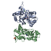
Mass: 65.409 Da / Num. of mol.: 5 / Source method: obtained synthetically / Formula: Zn
#3: Chemical ChemComp-CG8 / (2~{S})-2-azanyl-3-[6-methyl-2,4-bis(oxidanylidene)-5,7-dihydropyrrolo[3,4-d]pyrimidin-1-yl]propanoic acid


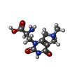
Mass: 254.243 Da / Num. of mol.: 2 / Source method: obtained synthetically / Formula: C10H14N4O4
#4: Chemical ChemComp-CL / CHLORIDE ION


Mass: 35.453 Da / Num. of mol.: 3 / Source method: obtained synthetically / Formula: Cl
#5: Chemical ChemComp-SO4 / SULFATE ION


Mass: 96.063 Da / Num. of mol.: 1 / Source method: obtained synthetically / Formula: SO4
#6: Water ChemComp-HOH / water


Mass: 18.015 Da / Num. of mol.: 46 / Source method: isolated from a natural source / Formula: H2O

-
Experimental details

-
Experiment

ExperimentMethod: X-RAY DIFFRACTION / Number of used crystals: 1

-
Sample preparation

CrystalDensity Matthews: 2.76 Å3/Da / Density % sol: 55.43 %
Crystal growTemperature: 295 K / Method: vapor diffusion, hanging drop / pH: 8.4
Details: 20% PEG3350, 0.1M BisTrisPropane pH 8.4, 0.2M sodium sulfate, 5 mM zinc acetate

-
Data collection

DiffractionMean temperature: 100 K
Diffraction sourceSource: SYNCHROTRON / Site: MAX II / Beamline: I911-3 / Wavelength: 1 Å
DetectorType: MARMOSAIC 225 mm CCD / Detector: CCD / Date: May 14, 2013
RadiationProtocol: SINGLE WAVELENGTH / Monochromatic (M) / Laue (L): M / Scattering type: x-ray
Radiation wavelengthWavelength: 1 Å / Relative weight: 1
ReflectionResolution: 2.4→28.374 Å / Num. obs: 25952 / % possible obs: 99.9 % / Redundancy: 4.4 % / Biso Wilson estimate: 49.94 Å2 / Rpim(I) all: 0.027 / Rrim(I) all: 0.059 / Rsym value: 0.052 / Net I/av σ(I): 10.2 / Net I/σ(I): 15.8
Reflection shell

Diffraction-ID: 1

Resolution (Å)Redundancy (%)Rmerge(I) obsMean I/σ(I) obsNum. unique obsRpim(I) allRrim(I) allRsym value% possible all
2.4-2.534.50.395237490.2090.4470.395100
2.53-2.684.40.262.835310.1380.2950.26100
2.68-2.874.40.1684.632990.0880.190.168100
2.87-3.14.40.0987.631210.0510.1110.098100
3.1-3.394.40.06211.328670.0330.070.062100
3.39-3.794.40.04613.426080.0240.0520.046100
3.79-4.384.40.03816.523220.020.0430.038100
4.38-5.374.30.03517.119730.0180.0390.035100
5.37-7.594.20.03716.915740.020.0420.037100
7.59-28.3743.80.0318.39080.0170.0350.0397.7

-
Processing

Software
NameVersionClassification
SCALA3.3.20data scaling
PHENIXrefinement
PDB_EXTRACT3.22data extraction
XDSdata reduction
PHASERphasing
RefinementMethod to determine structure: MOLECULAR REPLACEMENT
Starting model: 4mh5
Resolution: 2.4→28.374 Å / SU ML: 0.26 / Cross valid method: THROUGHOUT / σ(F): 1.36 / Phase error: 25.07
RfactorNum. reflection% reflection
Rfree0.2216 1318 5.08 %
Rwork0.1954 --
obs0.1967 25932 99.94 %
Solvent computationShrinkage radii: 0.9 Å / VDW probe radii: 1.11 Å
Displacement parametersBiso max: 161.66 Å2 / Biso mean: 70.1164 Å2 / Biso min: 32.24 Å2
Refinement stepCycle: final / Resolution: 2.4→28.374 Å
ProteinNucleic acidLigandSolventTotal
Num. atoms4022 0 49 46 4117
Biso mean--69.53 52.18 -
Num. residues----506
Refine LS restraints
Refine-IDTypeDev idealNumber
X-RAY DIFFRACTIONf_bond_d0.0034143
X-RAY DIFFRACTIONf_angle_d0.5675585
X-RAY DIFFRACTIONf_chiral_restr0.042614
X-RAY DIFFRACTIONf_plane_restr0.004699
X-RAY DIFFRACTIONf_dihedral_angle_d16.9832494
Refine LS restraints NCS
Ens-IDDom-IDAuth asym-IDNumberRefine-IDRmsType
11A3024X-RAY DIFFRACTION9.518TORSIONAL
12B3024X-RAY DIFFRACTION9.518TORSIONAL
LS refinement shell

Refine-ID: X-RAY DIFFRACTION / Rfactor Rfree error: 0 / Total num. of bins used: 9 / % reflection obs: 100 %

Resolution (Å)Rfactor RfreeNum. reflection RfreeRfactor RworkNum. reflection RworkNum. reflection all
2.4-2.49610.29331380.289727042842
2.4961-2.60960.2721480.259426732821
2.6096-2.74710.25571220.248427162838
2.7471-2.9190.28691470.258527062853
2.919-3.14410.27911530.243827032856
3.1441-3.460.23041550.218227092864
3.46-3.95950.22611480.190727372885
3.9595-4.98420.17251450.14627612906
4.9842-28.37550.20211620.169329053067
Refinement TLS params.

Method: refined / Refine-ID: X-RAY DIFFRACTION

IDL112)L122)L132)L222)L232)L332)S11 (Å °)S12 (Å °)S13 (Å °)S21 (Å °)S22 (Å °)S23 (Å °)S31 (Å °)S32 (Å °)S33 (Å °)T112)T122)T132)T222)T232)T332)Origin x (Å)Origin y (Å)Origin z (Å)
12.0181-1.7308-0.91911.79180.17874.3375-0.17780.06632.1037-0.13430.34151.4253-0.7661-0.5834-0.15740.54920.2036-0.04010.535-0.02141.4148-52.4571-5.9388-10.5265
25.1011.3661.57411.88570.5032.88770.0530.86580.3552-0.47960.01250.251-0.00720.2322-0.06910.41350.1225-0.07280.46480.08040.4328-42.8389-21.8484-20.1047
33.51061.94024.73322.68541.26367.99830.35250.27930.443-0.3565-0.17780.69720.2359-0.5396-0.14230.40540.0523-0.17370.4670.00660.6088-55.061-29.0889-18.2632
44.70123.5235-0.72515.4711-1.59085.46310.2134-0.44620.6780.1333-0.18920.46870.0545-0.22540.01540.24640.04530.01250.2807-0.0720.3746-42.979-18.348-3.0471
53.73951.601-0.20453.40920.66346.0813-0.14570.0626-0.6172-0.1745-0.0587-0.69640.07490.78460.06960.35850.10970.06550.5003-0.0310.5609-8.119-38.2843-10.4871
64.9377-3.3961.61873.44571.03785.7647-0.5129-0.2389-1.5291-0.29020.2443-0.30841.40520.13220.05690.79180.01550.32970.4359-0.01891.1509-16.5727-52.6225-12.6022
75.58280.7932.93185.4421.15419.2561-0.22970.78350.2439-0.88980.29610.2095-0.6157-0.3623-0.13640.416-0.08230.01350.3396-0.04580.4928-23.1044-37.6808-30.8462
84.89311.21150.41066.55942.51446.5669-0.0940.9217-1.0112-0.40450.3169-0.79340.32360.2627-0.25330.5915-0.08080.06980.6293-0.2560.6437-18.6814-44.183-34.7331
96.33510.0993-1.5645.8617-2.82637.7438-0.1672-0.2083-0.13160.1210.08790.0183-0.1548-0.19870.03230.27620.00380.04410.3343-0.08550.3687-21.7281-34.9618-10.8778
Refinement TLS group
IDRefine-IDRefine TLS-IDSelection detailsAuth asym-IDAuth seq-ID
1X-RAY DIFFRACTION1chain 'A' and (resid 5 through 36 )A5 - 36
2X-RAY DIFFRACTION2chain 'A' and (resid 37 through 173 )A37 - 173
3X-RAY DIFFRACTION3chain 'A' and (resid 174 through 214 )A174 - 214
4X-RAY DIFFRACTION4chain 'A' and (resid 215 through 258 )A215 - 258
5X-RAY DIFFRACTION5chain 'B' and (resid 6 through 67 )B6 - 67
6X-RAY DIFFRACTION6chain 'B' and (resid 68 through 103 )B68 - 103
7X-RAY DIFFRACTION7chain 'B' and (resid 104 through 124 )B104 - 124
8X-RAY DIFFRACTION8chain 'B' and (resid 125 through 214 )B125 - 214
9X-RAY DIFFRACTION9chain 'B' and (resid 215 through 257 )B215 - 257

+
About Yorodumi

-
News

-
Feb 9, 2022. New format data for meta-information of EMDB entries

New format data for meta-information of EMDB entries

  • Version 3 of the EMDB header file is now the official format.
  • The previous official version 1.9 will be removed from the archive.

Related info.:EMDB header

External links:wwPDB to switch to version 3 of the EMDB data model

-
Aug 12, 2020. Covid-19 info

Covid-19 info

URL: https://pdbjlvh1.pdbj.org/emnavi/covid19.php

New page: Covid-19 featured information page in EM Navigator.

Related info.:Covid-19 info / Mar 5, 2020. Novel coronavirus structure data

+
Mar 5, 2020. Novel coronavirus structure data

Novel coronavirus structure data

Related info.:Yorodumi Speices / Aug 12, 2020. Covid-19 info

External links:COVID-19 featured content - PDBj / Molecule of the Month (242):Coronavirus Proteases

+
Jan 31, 2019. EMDB accession codes are about to change! (news from PDBe EMDB page)

EMDB accession codes are about to change! (news from PDBe EMDB page)

  • The allocation of 4 digits for EMDB accession codes will soon come to an end. Whilst these codes will remain in use, new EMDB accession codes will include an additional digit and will expand incrementally as the available range of codes is exhausted. The current 4-digit format prefixed with “EMD-” (i.e. EMD-XXXX) will advance to a 5-digit format (i.e. EMD-XXXXX), and so on. It is currently estimated that the 4-digit codes will be depleted around Spring 2019, at which point the 5-digit format will come into force.
  • The EM Navigator/Yorodumi systems omit the EMD- prefix.

Related info.:Q: What is EMD? / ID/Accession-code notation in Yorodumi/EM Navigator

External links:EMDB Accession Codes are Changing Soon! / Contact to PDBj

+
Jul 12, 2017. Major update of PDB

Major update of PDB

  • wwPDB released updated PDB data conforming to the new PDBx/mmCIF dictionary.
  • This is a major update changing the version number from 4 to 5, and with Remediation, in which all the entries are updated.
  • In this update, many items about electron microscopy experimental information are reorganized (e.g. em_software).
  • Now, EM Navigator and Yorodumi are based on the updated data.

External links:wwPDB Remediation / Enriched Model Files Conforming to OneDep Data Standards Now Available in the PDB FTP Archive

-
Yorodumi

Thousand views of thousand structures

  • Yorodumi is a browser for structure data from EMDB, PDB, SASBDB, etc.
  • This page is also the successor to EM Navigator detail page, and also detail information page/front-end page for Omokage search.
  • The word "yorodu" (or yorozu) is an old Japanese word meaning "ten thousand". "mi" (miru) is to see.

Related info.:EMDB / PDB / SASBDB / Comparison of 3 databanks / Yorodumi Search / Aug 31, 2016. New EM Navigator & Yorodumi / Yorodumi Papers / Jmol/JSmol / Function and homology information / Changes in new EM Navigator and Yorodumi

Read more