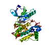
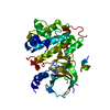
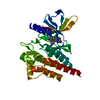
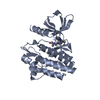
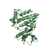
Entry Database : PDB / ID : 5y5uTitle Crystal structures of spleen tyrosine kinase in complex with a novel inhibitor Tyrosine-protein kinase SYK Keywords / / / / / / Function / homology Function Domain/homology Component
/ / / / / / / / / / / / / / / / / / / / / / / / / / / / / / / / / / / / / / / / / / / / / / / / / / / / / / / / / / / / / / / / / / / / / / / / / / / / / / / / / / / / / / / / / / / / / / / / / / / / / / / / / / / / / / / / / / / / / / / / / / / / / / / / / / / / / / / / / / / / / / / / Biological species Homo sapiens (human)Method / / / Resolution : 2.14 Å Authors Lee, S.J. / Lee, B.I. Journal : Mol. Cells / Year : 2018Title : Crystal Structures of Spleen Tyrosine Kinase in Complex with Two Novel 4-Aminopyrido[4,3-d] Pyrimidine Derivative Inhibitors.Authors : Lee, S.J. / Choi, J.S. / Bong, S.M. / Hwang, H.J. / Lee, J. / Song, H.J. / Lee, J. / Kim, J.H. / Koh, J.S. / Lee, B.I. History Deposition Aug 9, 2017 Deposition site / Processing site Revision 1.0 Jun 27, 2018 Provider / Type Revision 1.1 Jul 11, 2018 Group / Database references / Category Item / _citation.page_first / _citation.page_lastRevision 1.2 Nov 22, 2023 Group / Database references / Refinement descriptionCategory chem_comp_atom / chem_comp_bond ... chem_comp_atom / chem_comp_bond / database_2 / pdbx_initial_refinement_model Item / _database_2.pdbx_database_accession
Show all Show less
 Movie
Movie Controller

 Yorodumi
 Open data
 Basic information
 Components
 Keywords
 Function and homology information
 Homo sapiens (human)
 X-RAY DIFFRACTION /
  Authors
 Citation
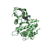
 Structure visualization
 Downloads & links
 Other downloads
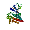


 PDBj
PDBj


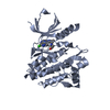
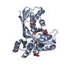
 Assembly
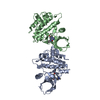


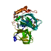
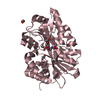
 Spodoptera frugiperda (fall armyworm)
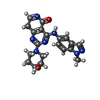
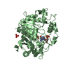
 Mass: 391.426 Da / Num. of mol.: 2 / Source method: obtained synthetically / Formula: C20H21N7O2
 Mass: 18.015 Da / Num. of mol.: 67 / Source method: isolated from a natural source / Formula: H2O
 Sample preparation
 / Beamline: BL26B1 / Wavelength: 1 Å
 Processing