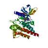
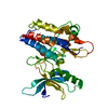
[English] 日本語
Yorodumi
- PDB-3bel: X-ray structure of EGFR in complex with oxime inhibitor -

+
Open data


ID or keywords:

Loading...

-
Basic information

Entry
Database: PDB / ID: 3bel
TitleX-ray structure of EGFR in complex with oxime inhibitor
ComponentsEpidermal growth factor receptor
KeywordsTRANSFERASE / kinase domain / Alternative splicing / Anti-oncogene / ATP-binding / Cell cycle / Disease mutation / Glycoprotein / Membrane / Nucleotide-binding / Phosphoprotein / Polymorphism / Receptor / Secreted / Transmembrane / Tyrosine-protein kinase / Ubl conjugation
Function / homology
Function and homology information


response to hydroxyisoflavone / multivesicular body, internal vesicle lumen / positive regulation of prolactin secretion / negative regulation of cardiocyte differentiation / positive regulation of protein kinase C activity / diterpenoid metabolic process / Shc-EGFR complex / ovulation cycle / Inhibition of Signaling by Overexpressed EGFR / epidermal growth factor receptor activity ...response to hydroxyisoflavone / multivesicular body, internal vesicle lumen / positive regulation of prolactin secretion / negative regulation of cardiocyte differentiation / positive regulation of protein kinase C activity / diterpenoid metabolic process / Shc-EGFR complex / ovulation cycle / Inhibition of Signaling by Overexpressed EGFR / epidermal growth factor receptor activity / EGFR interacts with phospholipase C-gamma / positive regulation of mucus secretion / response to UV-A / epidermal growth factor binding / PLCG1 events in ERBB2 signaling / tongue development / midgut development / ERBB2-EGFR signaling pathway / hydrogen peroxide metabolic process / PTK6 promotes HIF1A stabilization / digestive tract morphogenesis / morphogenesis of an epithelial fold / ERBB2 Activates PTK6 Signaling / intracellular vesicle / Signaling by EGFR / response to cobalamin / transmembrane receptor protein tyrosine kinase activator activity / protein tyrosine kinase activator activity / negative regulation of epidermal growth factor receptor signaling pathway / Signaling by ERBB4 / eyelid development in camera-type eye / regulation of phosphatidylinositol 3-kinase/protein kinase B signal transduction / protein insertion into membrane / cerebral cortex cell migration / ERBB2 Regulates Cell Motility / Respiratory syncytial virus (RSV) attachment and entry / regulation of JNK cascade / : / PI3K events in ERBB2 signaling / positive regulation of cyclin-dependent protein serine/threonine kinase activity / negative regulation of mitotic cell cycle / hair follicle development / Estrogen-dependent nuclear events downstream of ESR-membrane signaling / MAP kinase kinase kinase activity / embryonic placenta development / positive regulation of bone resorption / positive regulation of G1/S transition of mitotic cell cycle / GAB1 signalosome / salivary gland morphogenesis / peptidyl-tyrosine autophosphorylation / regulation of peptidyl-tyrosine phosphorylation / positive regulation of phosphorylation / positive regulation of glial cell proliferation / positive regulation of vasoconstriction / Signaling by ERBB2 / cellular response to epidermal growth factor stimulus / GRB2 events in EGFR signaling / SHC1 events in EGFR signaling / cellular response to cadmium ion / EGFR Transactivation by Gastrin / positive regulation of DNA repair / GRB2 events in ERBB2 signaling / TFAP2 (AP-2) family regulates transcription of growth factors and their receptors / transmembrane receptor protein tyrosine kinase activity / SHC1 events in ERBB2 signaling / positive regulation of synaptic transmission, glutamatergic / cellular response to dexamethasone stimulus / ossification / neurogenesis / basal plasma membrane / regulation of ERK1 and ERK2 cascade / neuron projection morphogenesis / positive regulation of superoxide anion generation / epithelial cell proliferation / positive regulation of DNA replication / Signal transduction by L1 / cellular response to estradiol stimulus / positive regulation of epithelial cell proliferation / NOTCH3 Activation and Transmission of Signal to the Nucleus / astrocyte activation / liver regeneration / positive regulation of protein localization to plasma membrane / EGFR downregulation / cellular response to amino acid stimulus / positive regulation of smooth muscle cell proliferation / Signaling by ERBB2 TMD/JMD mutants / positive regulation of MAP kinase activity / lung development / clathrin-coated endocytic vesicle membrane / Constitutive Signaling by EGFRvIII / Signaling by ERBB2 ECD mutants / Signaling by ERBB2 KD Mutants / epidermal growth factor receptor signaling pathway / negative regulation of protein catabolic process / receptor protein-tyrosine kinase / Downregulation of ERBB2 signaling / kinase binding / ruffle membrane / positive regulation of miRNA transcription / cell-cell adhesion
Similarity search - Function
: / Epidermal growth factor receptor transmembrane-juxtamembrane segment / Tyrosine protein kinase, EGF/ERB/XmrK receptor / Growth factor receptor domain 4 / Growth factor receptor domain IV / Receptor L-domain / Furin-like cysteine-rich domain / Receptor L-domain superfamily / Furin-like cysteine rich region / Receptor L domain ...: / Epidermal growth factor receptor transmembrane-juxtamembrane segment / Tyrosine protein kinase, EGF/ERB/XmrK receptor / Growth factor receptor domain 4 / Growth factor receptor domain IV / Receptor L-domain / Furin-like cysteine-rich domain / Receptor L-domain superfamily / Furin-like cysteine rich region / Receptor L domain / Furin-like repeat / Furin-like repeats / Growth factor receptor cysteine-rich domain superfamily / Tyrosine-protein kinase, catalytic domain / Tyrosine kinase, catalytic domain / Tyrosine protein kinases specific active-site signature. / Tyrosine-protein kinase, active site / Protein tyrosine and serine/threonine kinase / Serine-threonine/tyrosine-protein kinase, catalytic domain / Transferase(Phosphotransferase) domain 1 / Transferase(Phosphotransferase); domain 1 / Phosphorylase Kinase; domain 1 / Phosphorylase Kinase; domain 1 / Protein kinase, ATP binding site / Protein kinases ATP-binding region signature. / Protein kinase domain profile. / Protein kinase domain / Protein kinase-like domain superfamily / 2-Layer Sandwich / Orthogonal Bundle / Mainly Alpha / Alpha Beta
Similarity search - Domain/homology
PHOSPHATE ION / Chem-POX / Epidermal growth factor receptor
Similarity search - Component
Biological speciesHomo sapiens (human)
MethodX-RAY DIFFRACTION / SYNCHROTRON / MOLECULAR REPLACEMENT / molecular replacement / Resolution: 2.3 Å
AuthorsAbad, M.C. / Xu, G. / Neeper, M.P. / Struble, G.T. / Gaul, M.D. / Connolly, P.J.
CitationJournal: Bioorg.Med.Chem.Lett. / Year: 2008
Title: Discovery of novel 4-amino-6-arylaminopyrimidine-5-carbaldehyde oximes as dual inhibitors of EGFR and ErbB-2 protein tyrosine kinases.
Authors: Xu, G. / Searle, L.L. / Hughes, T.V. / Beck, A.K. / Connolly, P.J. / Abad, M.C. / Neeper, M.P. / Struble, G.T. / Springer, B.A. / Emanuel, S.L. / Gruninger, R.H. / Pandey, N. / Adams, M. / ...Authors: Xu, G. / Searle, L.L. / Hughes, T.V. / Beck, A.K. / Connolly, P.J. / Abad, M.C. / Neeper, M.P. / Struble, G.T. / Springer, B.A. / Emanuel, S.L. / Gruninger, R.H. / Pandey, N. / Adams, M. / Moreno-Mazza, S. / Fuentes-Pesquera, A.R. / Middleton, S.A. / Greenberger, L.M.
History
DepositionNov 19, 2007Deposition site: RCSB / Processing site: RCSB
Revision 1.0Jul 1, 2008Provider: repository / Type: Initial release
Revision 1.1Jul 13, 2011Group: Version format compliance
Revision 1.2Oct 25, 2017Group: Refinement description / Category: software
Revision 1.3Aug 30, 2023Group: Data collection / Database references ...Data collection / Database references / Derived calculations / Refinement description
Category: chem_comp_atom / chem_comp_bond ...chem_comp_atom / chem_comp_bond / database_2 / pdbx_initial_refinement_model / struct_site
Item: _database_2.pdbx_DOI / _database_2.pdbx_database_accession ..._database_2.pdbx_DOI / _database_2.pdbx_database_accession / _struct_site.pdbx_auth_asym_id / _struct_site.pdbx_auth_comp_id / _struct_site.pdbx_auth_seq_id

-
Structure visualization

Structure viewerMolecule:
MolmilJmol/JSmol

Downloads & links

-
Assembly

Deposited unit
A: Epidermal growth factor receptor
hetero molecules


Theoretical massNumber of molelcules
Total (without water)36,6964
Polymers36,0711
Non-polymers6253
Water48627
1


  • Idetical with deposited unit
  • defined by author&software
TypeNameSymmetry operationNumber
identity operation1_555x,y,z1
MethodPISA
Unit cell
Length a, b, c (Å)45.499, 67.686, 103.252
Angle α, β, γ (deg.)90.000, 90.000, 90.000
Int Tables number19
Space group name H-MP212121

-
Components

#1: Protein Epidermal growth factor receptor / Receptor tyrosine-protein kinase ErbB-1


Mass: 36070.809 Da / Num. of mol.: 1 / Fragment: Residues 702-1016
Source method: isolated from a genetically manipulated source
Details: Expressed open reading frame of construct is MHHHHHHVDLVPRGSHMA-(EGFR1 S671-G998
Source: (gene. exp.) Homo sapiens (human) / Strain: human strains undefined / Gene: EGFR, ERBB1 / Plasmid: modified pDEST8 / Production host: Spodoptera frugiperda (fall armyworm) / Strain (production host): sf9
References: UniProt: P00533, receptor protein-tyrosine kinase
#2: Chemical ChemComp-PO4 / PHOSPHATE ION


Mass: 94.971 Da / Num. of mol.: 2 / Source method: obtained synthetically / Formula: PO4
#3: Chemical ChemComp-POX / 4-amino-6-{[1-(3-fluorobenzyl)-1H-indazol-5-yl]amino}pyrimidine-5-carbaldehyde O-(2-methoxyethyl)oxime


Mass: 435.454 Da / Num. of mol.: 1 / Source method: obtained synthetically / Formula: C22H22FN7O2
#4: Water ChemComp-HOH / water


Mass: 18.015 Da / Num. of mol.: 27 / Source method: isolated from a natural source / Formula: H2O

-
Experimental details

-
Experiment

ExperimentMethod: X-RAY DIFFRACTION / Number of used crystals: 1

-
Sample preparation

CrystalDensity Matthews: 2.2 Å3/Da / Density % sol: 44.19 %
Crystal growTemperature: 295 K / Method: vapor diffusion, hanging drop / pH: 9
Details: 2M Na/K3PO4, 0.1M Caps pH 9.0 and 0.2M Li2SO4, VAPOR DIFFUSION, HANGING DROP, temperature 295K

-
Data collection

DiffractionMean temperature: 200 K
Diffraction sourceSource: SYNCHROTRON / Site: APS / Beamline: 14-ID-B / Wavelength: 1 Å
DetectorType: ADSC QUANTUM 210 / Detector: CCD / Date: Mar 16, 2005
RadiationProtocol: SINGLE WAVELENGTH / Monochromatic (M) / Laue (L): M / Scattering type: x-ray
Radiation wavelengthWavelength: 1 Å / Relative weight: 1
ReflectionResolution: 2.3→50 Å / Num. obs: 14413 / % possible obs: 97.1 % / Redundancy: 4.6 % / Biso Wilson estimate: 33 Å2 / Rmerge(I) obs: 0.108 / Χ2: 1.413 / Net I/σ(I): 14.8
Reflection shell
Resolution (Å)Redundancy (%)Rmerge(I) obsNum. unique allΧ2% possible all
2.3-2.384.80.18714451.353100
2.38-2.484.80.17814591.466100
2.48-2.594.80.15314521.399100
2.59-2.734.80.13214551.505100
2.73-2.94.80.12314591.31199.9
2.9-3.124.80.1114641.42999.7
3.12-3.444.70.09914591.4998.6
3.44-3.934.30.114241.42995.2
3.93-4.9540.09613661.33989.9
4.95-504.10.10714301.39388.5

-
Phasing

PhasingMethod: molecular replacement

-
Processing

Software
NameVersionClassificationNB
DENZOdata reduction
SCALEPACKdata scaling
CNSrefinement
PDB_EXTRACT3data extraction
HKL-2000data collection
CNSphasing
RefinementMethod to determine structure: MOLECULAR REPLACEMENT
Starting model: pdb id 2RGP
Resolution: 2.3→50 Å / Isotropic thermal model: anisotropic / Cross valid method: THROUGHOUT / σ(F): 0 / σ(I): 0
RfactorNum. reflection% reflectionSelection details
Rfree0.281 1455 9.9 %random
Rwork0.241 ---
obs-14321 97 %-
Displacement parametersBiso mean: 47.543 Å2
Baniso -1Baniso -2Baniso -3
1--11.488 Å20 Å20 Å2
2--5.989 Å20 Å2
3---5.499 Å2
Refinement stepCycle: LAST / Resolution: 2.3→50 Å
ProteinNucleic acidLigandSolventTotal
Num. atoms2250 0 42 27 2319
Refine LS restraints
Refine-IDTypeDev idealDev ideal target
X-RAY DIFFRACTIONc_mcbond_it1.5861.5
X-RAY DIFFRACTIONc_scbond_it2.2752
X-RAY DIFFRACTIONc_mcangle_it2.7232
X-RAY DIFFRACTIONc_scangle_it3.3822.5
Xplor file
Refine-IDSerial noParam file
X-RAY DIFFRACTION1MSI_CNX_TOPPAR:protein_rep.param
X-RAY DIFFRACTION2inh.par
X-RAY DIFFRACTION3MSI_CNX_TOPPAR:water_rep.param
X-RAY DIFFRACTION4MSI_CNX_TOPPAR:ion.param

+
About Yorodumi

-
News

-
Feb 9, 2022. New format data for meta-information of EMDB entries

New format data for meta-information of EMDB entries

  • Version 3 of the EMDB header file is now the official format.
  • The previous official version 1.9 will be removed from the archive.

Related info.:EMDB header

External links:wwPDB to switch to version 3 of the EMDB data model

-
Aug 12, 2020. Covid-19 info

Covid-19 info

URL: https://pdbjlvh1.pdbj.org/emnavi/covid19.php

New page: Covid-19 featured information page in EM Navigator.

Related info.:Covid-19 info / Mar 5, 2020. Novel coronavirus structure data

+
Mar 5, 2020. Novel coronavirus structure data

Novel coronavirus structure data

Related info.:Yorodumi Speices / Aug 12, 2020. Covid-19 info

External links:COVID-19 featured content - PDBj / Molecule of the Month (242):Coronavirus Proteases

+
Jan 31, 2019. EMDB accession codes are about to change! (news from PDBe EMDB page)

EMDB accession codes are about to change! (news from PDBe EMDB page)

  • The allocation of 4 digits for EMDB accession codes will soon come to an end. Whilst these codes will remain in use, new EMDB accession codes will include an additional digit and will expand incrementally as the available range of codes is exhausted. The current 4-digit format prefixed with “EMD-” (i.e. EMD-XXXX) will advance to a 5-digit format (i.e. EMD-XXXXX), and so on. It is currently estimated that the 4-digit codes will be depleted around Spring 2019, at which point the 5-digit format will come into force.
  • The EM Navigator/Yorodumi systems omit the EMD- prefix.

Related info.:Q: What is EMD? / ID/Accession-code notation in Yorodumi/EM Navigator

External links:EMDB Accession Codes are Changing Soon! / Contact to PDBj

+
Jul 12, 2017. Major update of PDB

Major update of PDB

  • wwPDB released updated PDB data conforming to the new PDBx/mmCIF dictionary.
  • This is a major update changing the version number from 4 to 5, and with Remediation, in which all the entries are updated.
  • In this update, many items about electron microscopy experimental information are reorganized (e.g. em_software).
  • Now, EM Navigator and Yorodumi are based on the updated data.

External links:wwPDB Remediation / Enriched Model Files Conforming to OneDep Data Standards Now Available in the PDB FTP Archive

-
Yorodumi

Thousand views of thousand structures

  • Yorodumi is a browser for structure data from EMDB, PDB, SASBDB, etc.
  • This page is also the successor to EM Navigator detail page, and also detail information page/front-end page for Omokage search.
  • The word "yorodu" (or yorozu) is an old Japanese word meaning "ten thousand". "mi" (miru) is to see.

Related info.:EMDB / PDB / SASBDB / Comparison of 3 databanks / Yorodumi Search / Aug 31, 2016. New EM Navigator & Yorodumi / Yorodumi Papers / Jmol/JSmol / Function and homology information / Changes in new EM Navigator and Yorodumi

Read more