 Movie
Movie Controller

[English] 日本語
 Yorodumi
- PDB-1uc0: Crystal structure of wild-type hen-egg white lysozyme singly labe... -
+ Open data

- Basic information
| Entry | Database: PDB / ID: 1uc0 | |||||||||
|---|---|---|---|---|---|---|---|---|---|---|
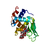
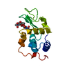
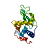
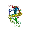
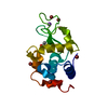
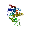
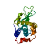
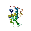
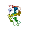
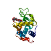
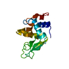
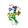
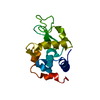
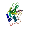
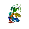
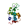
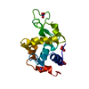
| Title | Crystal structure of wild-type hen-egg white lysozyme singly labeled with 2',3'-epoxypropyl beta-glycoside of N-acetyllactosamine | |||||||||
 Components Components | Lysozyme C | |||||||||
 Keywords Keywords | HYDROLASE / Protein-carbohydrate complex | |||||||||
| Function / homology |  Function and homology information Function and homology informationLactose synthesis / Antimicrobial peptides / Neutrophil degranulation / beta-N-acetylglucosaminidase activity / cell wall macromolecule catabolic process / lysozyme / lysozyme activity / killing of cells of another organism / defense response to Gram-negative bacterium / defense response to bacterium ...Lactose synthesis / Antimicrobial peptides / Neutrophil degranulation / beta-N-acetylglucosaminidase activity / cell wall macromolecule catabolic process / lysozyme / lysozyme activity / killing of cells of another organism / defense response to Gram-negative bacterium / defense response to bacterium / defense response to Gram-positive bacterium / endoplasmic reticulum / : / identical protein binding / cytoplasm Similarity search - Function | |||||||||
| Biological species |  | |||||||||
| Method |  X-RAY DIFFRACTION / MOLECULAR REPLACEMENT / Resolution: 1.85 Å X-RAY DIFFRACTION / MOLECULAR REPLACEMENT / Resolution: 1.85 Å | |||||||||
 Authors Authors | Muraki, M. / Harata, K. | |||||||||
 Citation Citation | Journal: J.MOL.RECOG. / Year: 2003 Title: X-ray structural analysis of the ligand-recognition mechanism in the dual-affinity labeling of c-type lysozyme with 2',3'-epoxypropyl beta-glycoside of N-acetyllactosamine Authors: Muraki, M. / Harata, K. | |||||||||
| History |
|
- Structure visualization
| Structure viewer | Molecule: MolmilJmol/JSmol |
|---|
- Downloads & links
-Download
| PDBx/mmCIF format | 1uc0.cif.gz | 41.2 KB | Display | PDBx/mmCIF format |
|---|---|---|---|---|
| PDB format | pdb1uc0.ent.gz | 27 KB | Display | PDB format |
| PDBx/mmJSON format | 1uc0.json.gz | Tree view | PDBx/mmJSON format | |
| Others |  Other downloads Other downloads |
-Validation report
| Arichive directory | https://data.pdbj.org/pub/pdb/validation_reports/uc/1uc0ftp://data.pdbj.org/pub/pdb/validation_reports/uc/1uc0 | HTTPS FTP |
|---|
-Related structure data


-Links
 PDBj
PDBj


- Assembly
| Deposited unit | 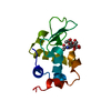
| ||||||||
|---|---|---|---|---|---|---|---|---|---|
| 1 |
| ||||||||
| Unit cell |
|
-Components
| #1: Protein | Mass: 14331.160 Da / Num. of mol.: 1 / Source method: isolated from a natural source / Source: (natural) |
|---|---|
| #2: Polysaccharide | beta-D-galactopyranose-(1-4)-2-acetamido-2-deoxy-beta-D-glucopyranose Source method: isolated from a genetically manipulated source |
| #3: Chemical | ChemComp-GOL /   Mass: 92.094 Da / Num. of mol.: 1 / Source method: obtained synthetically / Formula: C3H8O3 Mass: 92.094 Da / Num. of mol.: 1 / Source method: obtained synthetically / Formula: C3H8O3 |
| #4: Water | ChemComp-HOH /  Mass: 18.015 Da / Num. of mol.: 95 / Source method: isolated from a natural source / Formula: H2O Mass: 18.015 Da / Num. of mol.: 95 / Source method: isolated from a natural source / Formula: H2O |
| Has protein modification | Y |
-Experimental details
-Experiment
| Experiment | Method: X-RAY DIFFRACTION / Number of used crystals: 1 |
|---|
- Sample preparation
| Crystal | Density Matthews: 1.88 Å3/Da / Density % sol: 34.1 % |
|---|---|
| Crystal grow | Temperature: 298 K / Method: vapor diffusion, sitting drop / pH: 4.5 Details: sodium acetate, sodium chloride, pH 4.5, VAPOR DIFFUSION, SITTING DROP, temperature 298.0K |
| Crystal grow | *PLUS Method: vapor diffusion / Details: used microseeding |
| Components of the solutions | *PLUS Conc.: 50 mM / Common name: sodium acetate / Details: pH4.5 |
-Data collection
| Diffraction | Mean temperature: 283 K |
|---|---|
| Diffraction source | Source: ROTATING ANODE / Type: ENRAF-NONIUS FR571 / Wavelength: 1.5418 Å |
| Detector | Type: ENRAF-NONIUS FAST / Detector: AREA DETECTOR |
| Radiation | Protocol: SINGLE WAVELENGTH / Monochromatic (M) / Laue (L): M / Scattering type: x-ray |
| Radiation wavelength | Wavelength: 1.5418 Å / Relative weight: 1 |
| Reflection | Resolution: 1.78→27.6 Å / Num. obs: 74009 / % possible obs: 90.8 % / Observed criterion σ(I): 0 / Redundancy: 6.8 % / Rmerge(I) obs: 0.048 |
| Reflection shell | Resolution: 1.85→1.89 Å / Rmerge(I) obs: 0.258 |
| Reflection | *PLUS Num. obs: 10897 / Redundancy: 6.8 % / Num. measured all: 74099 |
- Processing
| Software |
| ||||||||||||||||||||||||
|---|---|---|---|---|---|---|---|---|---|---|---|---|---|---|---|---|---|---|---|---|---|---|---|---|---|
| Refinement | Method to determine structure: MOLECULAR REPLACEMENT Starting model: Wild-type Hen-egg white lysozyme Resolution: 1.85→8 Å / σ(F): 2
| ||||||||||||||||||||||||
| Displacement parameters | Biso mean: 14.89 Å2 | ||||||||||||||||||||||||
| Refinement step | Cycle: LAST / Resolution: 1.85→8 Å
| ||||||||||||||||||||||||
| Refine LS restraints |
| ||||||||||||||||||||||||
| Refinement | *PLUS Rfactor Rwork: 0.19 | ||||||||||||||||||||||||
| Solvent computation | *PLUS | ||||||||||||||||||||||||
| Displacement parameters | *PLUS | ||||||||||||||||||||||||
| Refine LS restraints | *PLUS
|
