 Movie
Movie Controller

[English] 日本語
 Yorodumi
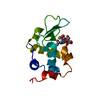
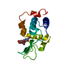
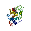
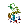
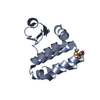
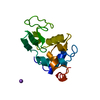
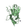
- PDB-1ubz: Crystal structure of Glu102-mutant human lysozyme doubly labeled ... -
+ Open data

- Basic information
| Entry | Database: PDB / ID: 1ubz | |||||||||
|---|---|---|---|---|---|---|---|---|---|---|
| Title | Crystal structure of Glu102-mutant human lysozyme doubly labeled with 2',3'-epoxypropyl beta-glycoside of N-acetyllactosamine | |||||||||
 Components Components | Lysozyme C | |||||||||
 Keywords Keywords | HYDROLASE / Protein-Carbohydrate complex | |||||||||
| Function / homology |  Function and homology information Function and homology informationmetabolic process / cytolysis / antimicrobial humoral response / retina homeostasis / Antimicrobial peptides / specific granule lumen / lysozyme / lysozyme activity / azurophil granule lumen / tertiary granule lumen ...metabolic process / cytolysis / antimicrobial humoral response / retina homeostasis / Antimicrobial peptides / specific granule lumen / lysozyme / lysozyme activity / azurophil granule lumen / tertiary granule lumen / killing of cells of another organism / defense response to Gram-negative bacterium / defense response to bacterium / defense response to Gram-positive bacterium / Amyloid fiber formation / inflammatory response / Neutrophil degranulation / : / extracellular exosome / extracellular region / identical protein binding Similarity search - Function | |||||||||
| Biological species |  Homo sapiens (human) Homo sapiens (human) | |||||||||
| Method |  X-RAY DIFFRACTION / SYNCHROTRON / MOLECULAR REPLACEMENT / Resolution: 2 Å X-RAY DIFFRACTION / SYNCHROTRON / MOLECULAR REPLACEMENT / Resolution: 2 Å | |||||||||
 Authors Authors | Muraki, M. / Harata, K. | |||||||||
 Citation Citation | Journal: J.MOL.RECOG. / Year: 2003 Title: X-ray structural analysis of the ligand-recognition mechanism in the dual-affinity labeling of c-type lysozyme with 2',3'-epoxypropyl beta-glycoside of N-acetyllactosamine Authors: Muraki, M. / Harata, K. | |||||||||
| History |
|
- Structure visualization
| Structure viewer | Molecule: MolmilJmol/JSmol |
|---|
- Downloads & links
-Download
| PDBx/mmCIF format | 1ubz.cif.gz | 43 KB | Display | PDBx/mmCIF format |
|---|---|---|---|---|
| PDB format | pdb1ubz.ent.gz | 28.5 KB | Display | PDB format |
| PDBx/mmJSON format | 1ubz.json.gz | Tree view | PDBx/mmJSON format | |
| Others |  Other downloads Other downloads |
-Validation report
| Arichive directory | https://data.pdbj.org/pub/pdb/validation_reports/ub/1ubzftp://data.pdbj.org/pub/pdb/validation_reports/ub/1ubz | HTTPS FTP |
|---|
-Related structure data


-Links
 PDBj
PDBj

- Assembly
| Deposited unit | 
| ||||||||
|---|---|---|---|---|---|---|---|---|---|
| 1 |
| ||||||||
| Unit cell |
|
-Components
| #1: Protein | Mass: 14734.719 Da / Num. of mol.: 1 / Mutation: D102E Source method: isolated from a genetically manipulated source Source: (gene. exp.) Homo sapiens (human) / Plasmid: YEp51 / Production host:  | ||||||
|---|---|---|---|---|---|---|---|
| #2: Polysaccharide | Source method: isolated from a genetically manipulated source #3: Chemical |   Mass: 92.094 Da / Num. of mol.: 2 / Source method: obtained synthetically / Formula: C3H8O3 Mass: 92.094 Da / Num. of mol.: 2 / Source method: obtained synthetically / Formula: C3H8O3#4: Water | ChemComp-HOH / |  Mass: 18.015 Da / Num. of mol.: 113 / Source method: isolated from a natural source / Formula: H2O Mass: 18.015 Da / Num. of mol.: 113 / Source method: isolated from a natural source / Formula: H2OHas protein modification | Y | |
-Experimental details
-Experiment
| Experiment | Method: X-RAY DIFFRACTION / Number of used crystals: 1 |
|---|
- Sample preparation
| Crystal | Density Matthews: 1.67 Å3/Da / Density % sol: 25.7 % |
|---|---|
| Crystal grow | Temperature: 298 K / Method: vapor diffusion, sitting drop / pH: 4.5 Details: sodium acetate, sodium nitrate, pH 4.5, VAPOR DIFFUSION, SITTING DROP, temperature 298.0K |
| Crystal grow | *PLUS Method: vapor diffusion / Details: used microseeding |
| Components of the solutions | *PLUS Conc.: 50 mM / Common name: sodium acetate / Details: pH4.5 |
-Data collection
| Diffraction | Mean temperature: 100 K |
|---|---|
| Diffraction source | Source: SYNCHROTRON / Site: Photon Factory  / Beamline: BL-6A / Wavelength: 1 Å / Beamline: BL-6A / Wavelength: 1 Å |
| Detector | Type: ADSC QUANTUM 4 / Detector: CCD / Date: Jun 16, 2000 |
| Radiation | Protocol: SINGLE WAVELENGTH / Monochromatic (M) / Laue (L): M / Scattering type: x-ray |
| Radiation wavelength | Wavelength: 1 Å / Relative weight: 1 |
| Reflection | Resolution: 2→28.8 Å / Num. obs: 41274 / % possible obs: 87.6 % / Observed criterion σ(I): 0 / Redundancy: 5.26 % / Rmerge(I) obs: 0.076 |
| Reflection shell | Resolution: 2→2.03 Å / % possible all: 69 |
| Reflection | *PLUS Num. obs: 7845 / Num. measured all: 41274 |
| Reflection shell | *PLUS Lowest resolution: 2.07 Å / Rmerge(I) obs: 0.15 |
- Processing
| Software |
| |||||||||||||||||||||
|---|---|---|---|---|---|---|---|---|---|---|---|---|---|---|---|---|---|---|---|---|---|---|
| Refinement | Method to determine structure: MOLECULAR REPLACEMENT / Resolution: 2→8 Å / σ(F): 2
| |||||||||||||||||||||
| Displacement parameters | Biso mean: 8.3 Å2 | |||||||||||||||||||||
| Refinement step | Cycle: LAST / Resolution: 2→8 Å
| |||||||||||||||||||||
| Refine LS restraints |
| |||||||||||||||||||||
| Refine LS restraints | *PLUS
|
