 Movie
Movie Controller


+ Open data

- Basic information
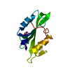
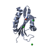
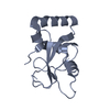
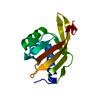
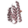
| Entry | Database: PDB / ID: 1t1j | ||||||
|---|---|---|---|---|---|---|---|
| Title | Crystal structure of genomics APC5043 | ||||||
 Components Components | hypothetical protein | ||||||
 Keywords Keywords | structural genomics / unknown function / PSI / Protein Structure Initiative / Midwest Center for Structural Genomics / MCSG | ||||||
| Function / homology | Hypothetical protein PA1492 / Domain of unknown function DUF1937 / Domain of unknown function (DUF1937) / Rossmann fold / 3-Layer(aba) Sandwich / Alpha Beta / DUF1937 domain-containing protein Function and homology information Function and homology information | ||||||
| Biological species |   Pseudomonas aeruginosa (bacteria) Pseudomonas aeruginosa (bacteria) | ||||||
| Method |  X-RAY DIFFRACTION / SYNCHROTRON / SAD / Resolution: 1.7 Å X-RAY DIFFRACTION / SYNCHROTRON / SAD / Resolution: 1.7 Å | ||||||
 Authors Authors | Dong, A. / Xu, X. / Liu, Y. / Zhang, R. / Savchenko, A. / Edwards, A. / Midwest Center for Structural Genomics (MCSG) | ||||||
 Citation Citation | Journal: TO BE PUBLISHED Title: Crystal Structure of Conserved Hypothetical Protein PA1492 from Pseudomonas aeruginosa Authors: Dong, A. / Xu, X. / Liu, Y. / Zhang, R. / Savchenko, A. / Edwards, A. | ||||||
| History |
|
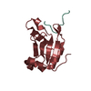
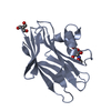
- Structure visualization
| Structure viewer | Molecule: MolmilJmol/JSmol |
|---|
- Downloads & links
-Download
| PDBx/mmCIF format | 1t1j.cif.gz | 63.4 KB | Display | PDBx/mmCIF format |
|---|---|---|---|---|
| PDB format | pdb1t1j.ent.gz | 46.7 KB | Display | PDB format |
| PDBx/mmJSON format | 1t1j.json.gz | Tree view | PDBx/mmJSON format | |
| Others |  Other downloads Other downloads |
-Validation report
| Arichive directory | https://data.pdbj.org/pub/pdb/validation_reports/t1/1t1jftp://data.pdbj.org/pub/pdb/validation_reports/t1/1t1j | HTTPS FTP |
|---|
-Related structure data
| Similar structure data | |
|---|---|
| Other databases |


-Links
 PDBj
PDBj- Assembly
| Deposited unit | 
| ||||||||
|---|---|---|---|---|---|---|---|---|---|
| 1 | 
| ||||||||
| 2 | 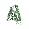
| ||||||||
| 3 | 
| ||||||||
| Unit cell |
|
-Components
| #1: Protein | Mass: 14367.354 Da / Num. of mol.: 2 Source method: isolated from a genetically manipulated source Source: (gene. exp.) Pseudomonas aeruginosa (bacteria) / Plasmid: pET15b / Species (production host): Escherichia coli / Production host: #2: Water | ChemComp-HOH / |  Mass: 18.015 Da / Num. of mol.: 146 / Source method: isolated from a natural source / Formula: H2O Mass: 18.015 Da / Num. of mol.: 146 / Source method: isolated from a natural source / Formula: H2O |
|---|
-Experimental details
-Experiment
| Experiment | Method: X-RAY DIFFRACTION / Number of used crystals: 1 |
|---|
- Sample preparation
| Crystal | Density Matthews: 2.76 Å3/Da / Density % sol: 55.1 % |
|---|---|
| Crystal grow | Temperature: 300 K / Method: vapor diffusion, hanging drop / pH: 8.5 Details: 3.5M Sodium formate, 4% Ethylene, pH 8.5, VAPOR DIFFUSION, HANGING DROP, temperature 300K |
-Data collection
| Diffraction | Mean temperature: 100 K |
|---|---|
| Diffraction source | Source: SYNCHROTRON / Site: APS  / Beamline: 19-BM / Wavelength: 0.96411 Å / Beamline: 19-BM / Wavelength: 0.96411 Å |
| Detector | Type: ADSC QUANTUM 4 / Detector: CCD / Date: Feb 28, 2004 |
| Radiation | Protocol: SINGLE WAVELENGTH / Monochromatic (M) / Laue (L): M / Scattering type: x-ray |
| Radiation wavelength | Wavelength: 0.96411 Å / Relative weight: 1 |
| Reflection | Resolution: 1.7→31.98 Å / Num. all: 63515 / Num. obs: 59550 / % possible obs: 93.8 % / Observed criterion σ(F): 0 / Observed criterion σ(I): -3 / Redundancy: 6.2 % / Biso Wilson estimate: 22.2 Å2 / Rmerge(I) obs: 0.052 / Rsym value: 0.052 / Net I/σ(I): 7.7 |
| Reflection shell | Resolution: 1.7→1.76 Å / Redundancy: 5.9 % / Rmerge(I) obs: 0.844 / Mean I/σ(I) obs: 1.42 / Num. unique all: 6351 / Rsym value: 0.844 / % possible all: 99.9 |
- Processing
| Software |
| ||||||||||||||||||||||||||||||||||||
|---|---|---|---|---|---|---|---|---|---|---|---|---|---|---|---|---|---|---|---|---|---|---|---|---|---|---|---|---|---|---|---|---|---|---|---|---|---|
| Refinement | Method to determine structure: SAD / Resolution: 1.7→31.98 Å / Rfactor Rfree error: 0.007 / Data cutoff high absF: 463098.63 / Data cutoff low absF: 0 / Isotropic thermal model: RESTRAINED / Cross valid method: maximum likelihood / σ(F): 0 / Stereochemistry target values: Engh & Huber
| ||||||||||||||||||||||||||||||||||||
| Solvent computation | Solvent model: FLAT MODEL / Bsol: 50.6097 Å2 / ksol: 0.410827 e/Å3 | ||||||||||||||||||||||||||||||||||||
| Displacement parameters | Biso mean: 27.9 Å2
| ||||||||||||||||||||||||||||||||||||
| Refine analyze |
| ||||||||||||||||||||||||||||||||||||
| Refinement step | Cycle: LAST / Resolution: 1.7→31.98 Å
| ||||||||||||||||||||||||||||||||||||
| Refine LS restraints |
| ||||||||||||||||||||||||||||||||||||
| LS refinement shell | Resolution: 1.7→1.81 Å / Rfactor Rfree error: 0.029 / Total num. of bins used: 6
| ||||||||||||||||||||||||||||||||||||
| Xplor file |
|
