 Movie
Movie Controller

[English] 日本語
 Yorodumi
- PDB-1dmn: CRYSTAL STRUCTURE OF MUTANT ENZYME Y32F/Y57F OF KETOSTEROID ISOME... -
+ Open data

- Basic information
| Entry | Database: PDB / ID: 1dmn | ||||||
|---|---|---|---|---|---|---|---|
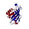
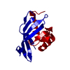
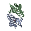
| Title | CRYSTAL STRUCTURE OF MUTANT ENZYME Y32F/Y57F OF KETOSTEROID ISOMERASE FROM PSEUDOMONAS PUTIDA BIOTYPE B | ||||||
 Components Components | STEROID DELTA-ISOMERASE | ||||||
 Keywords Keywords | ISOMERASE / CONESHELL / CLOSED BARREL / CURVED B-SHEET | ||||||
| Function / homology |  Function and homology information Function and homology informationsteroid Delta-isomerase / steroid Delta-isomerase activity / steroid metabolic process Similarity search - Function | ||||||
| Biological species |  Pseudomonas putida (bacteria) Pseudomonas putida (bacteria) | ||||||
| Method |  X-RAY DIFFRACTION / Resolution: 2.05 Å X-RAY DIFFRACTION / Resolution: 2.05 Å | ||||||
 Authors Authors | Kim, D.H. / Jang, D.S. / Nam, G.H. / Oh, B.H. / Choi, K.Y. | ||||||
 Citation Citation | Journal: Biochemistry / Year: 2000 Title: Contribution of the hydrogen-bond network involving a tyrosine triad in the active site to the structure and function of a highly proficient ketosteroid isomerase from Pseudomonas putida biotype B. Authors: Kim, D.H. / Jang, D.S. / Nam, G.H. / Choi, G. / Kim, J.S. / Ha, N.C. / Kim, M.S. / Oh, B.H. / Choi, K.Y. #1: Journal: Biochemistry / Year: 1997Title: High-Resolution Crystal Structures of delta(5)-3-Ketosteroid Isomerase with and without a Reaction Intermediate Analogue Authors: Kim, S.W. / Cha, S.S. / Cho, H.S. / Kim, J.S. / Ha, N.C. / Cho, M.J. / Joo, S. / Kim, K.K. / Choi, K.Y. / Oh, B.H. #2: Journal: Biochemistry / Year: 1998Title: Crystal Structure and Enzyme Mechanism of delta(5)-3-Ketosteroid Isomerase from Pseudomonas testosteroni Authors: Cho, H.S. / Choi, G. / Choi, K.Y. / Oh, B.H. #3: Journal: Biochemistry / Year: 1999Title: Crystal Structure of delta(5)-3-Ketosteroid Isomerase from Pseudomonas testosteroni in Complex with Equilenin Settles the Correct Hydrogen Bonding Scheme for Transition State Stabilization Authors: Cho, H.S. / Ha, N.C. / Choi, G. / Kim, H.J. / Lee, D. / Oh, K.S. / Kim, K.S. / Lee, W. / Choi, K.Y. / Oh, B.H. | ||||||
| History |
|
- Structure visualization
| Structure viewer | Molecule: MolmilJmol/JSmol |
|---|
- Downloads & links
-Download
| PDBx/mmCIF format | 1dmn.cif.gz | 37.4 KB | Display | PDBx/mmCIF format |
|---|---|---|---|---|
| PDB format | pdb1dmn.ent.gz | 25.6 KB | Display | PDB format |
| PDBx/mmJSON format | 1dmn.json.gz | Tree view | PDBx/mmJSON format | |
| Others |  Other downloads Other downloads |
-Validation report
| Arichive directory | https://data.pdbj.org/pub/pdb/validation_reports/dm/1dmnftp://data.pdbj.org/pub/pdb/validation_reports/dm/1dmn | HTTPS FTP |
|---|
-Related structure data


-Links
 PDBj
PDBj
- Assembly
| Deposited unit | 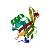
| ||||||||
|---|---|---|---|---|---|---|---|---|---|
| 1 | 
| ||||||||
| Unit cell |
|
-Components
| #1: Protein | Mass: 14516.501 Da / Num. of mol.: 1 / Mutation: Y32F, Y57F Source method: isolated from a genetically manipulated source Source: (gene. exp.) Pseudomonas putida (bacteria) / Plasmid: PKK223-3 / Production host: |
|---|---|
| #2: Water | ChemComp-HOH /  Mass: 18.015 Da / Num. of mol.: 48 / Source method: isolated from a natural source / Formula: H2O Mass: 18.015 Da / Num. of mol.: 48 / Source method: isolated from a natural source / Formula: H2O |
-Experimental details
-Experiment
| Experiment | Method: X-RAY DIFFRACTION / Number of used crystals: 1 |
|---|
- Sample preparation
| Crystal | Density Matthews: 2.25 Å3/Da / Density % sol: 45.39 % | |||||||||||||||
|---|---|---|---|---|---|---|---|---|---|---|---|---|---|---|---|---|
| Crystal grow | Temperature: 295 K / Method: vapor diffusion, hanging drop / pH: 4.6 Details: SODIUM ACETATE, AMMONIUM ACETATE, pH 4.6, VAPOR DIFFUSION, HANGING DROP, temperature 22K | |||||||||||||||
| Crystal grow | *PLUS | |||||||||||||||
| Components of the solutions | *PLUS
|
-Data collection
| Diffraction | Mean temperature: 295 K |
|---|---|
| Diffraction source | Source: ROTATING ANODE / Type: MACSCIENCE / Wavelength: 1.5418 |
| Detector | Type: MACSCIENCE / Detector: IMAGE PLATE / Date: Nov 20, 1999 |
| Radiation | Protocol: SINGLE WAVELENGTH / Monochromatic (M) / Laue (L): M / Scattering type: x-ray |
| Radiation wavelength | Wavelength: 1.5418 Å / Relative weight: 1 |
| Reflection | Resolution: 2.05→20 Å / Num. all: 9209 / Num. obs: 8592 / % possible obs: 93.3 % / Observed criterion σ(F): 0.5 / Observed criterion σ(I): 0.5 / Redundancy: 5.5 % / Biso Wilson estimate: 14.7 Å2 / Rmerge(I) obs: 0.066 / Net I/σ(I): 14.5 |
| Reflection shell | Resolution: 2.05→2.12 Å / Redundancy: 2.3 % / Rmerge(I) obs: 0.22 / % possible all: 89 |
- Processing
| Software |
| ||||||||||||||||||||||||||||||||||||||||||||||||||||||||||||
|---|---|---|---|---|---|---|---|---|---|---|---|---|---|---|---|---|---|---|---|---|---|---|---|---|---|---|---|---|---|---|---|---|---|---|---|---|---|---|---|---|---|---|---|---|---|---|---|---|---|---|---|---|---|---|---|---|---|---|---|---|---|
| Refinement | Resolution: 2.05→20 Å / σ(F): 0.5 / σ(I): 0.5 / Stereochemistry target values: ENGH & HUBER
| ||||||||||||||||||||||||||||||||||||||||||||||||||||||||||||
| Refinement step | Cycle: LAST / Resolution: 2.05→20 Å
| ||||||||||||||||||||||||||||||||||||||||||||||||||||||||||||
| Refine LS restraints |
| ||||||||||||||||||||||||||||||||||||||||||||||||||||||||||||
| Software | *PLUS Name: X-PLOR / Version: 3.843 / Classification: refinement | ||||||||||||||||||||||||||||||||||||||||||||||||||||||||||||
| Refinement | *PLUS Lowest resolution: 20 Å / σ(F): 0.5 / % reflection Rfree: 5 % | ||||||||||||||||||||||||||||||||||||||||||||||||||||||||||||
| Solvent computation | *PLUS | ||||||||||||||||||||||||||||||||||||||||||||||||||||||||||||
| Displacement parameters | *PLUS Biso mean: 25.2 Å2 |
