 Movie
Movie Controller

[English] 日本語
 Yorodumi
- PDB-3ipt: Crystal Structure of Ketosteroid Isomerase Y16S/D40N from Pseudom... -
+ Open data

- Basic information
| Entry | Database: PDB / ID: 3ipt | ||||||
|---|---|---|---|---|---|---|---|
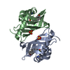
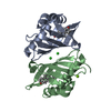
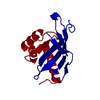
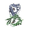
| Title | Crystal Structure of Ketosteroid Isomerase Y16S/D40N from Pseudomonas putida with Bound Equilenin | ||||||
 Components Components | Steroid Delta-isomerase | ||||||
 Keywords Keywords | ISOMERASE / enzyme-ligand complex / protein cavity / Lipid metabolism / Steroid metabolism | ||||||
| Function / homology |  Function and homology information Function and homology informationsteroid Delta-isomerase / steroid Delta-isomerase activity / steroid metabolic process Similarity search - Function | ||||||
| Biological species |  Pseudomonas putida (bacteria) Pseudomonas putida (bacteria) | ||||||
| Method |  X-RAY DIFFRACTION / SYNCHROTRON / MOLECULAR REPLACEMENT / Resolution: 1.632 Å X-RAY DIFFRACTION / SYNCHROTRON / MOLECULAR REPLACEMENT / Resolution: 1.632 Å | ||||||
 Authors Authors | Fenn, T.D. / Sigala, P.A. / Herschlag, D. | ||||||
 Citation Citation | Journal: Proc.Natl.Acad.Sci.USA / Year: 2010 Title: Dissecting the paradoxical effects of hydrogen bond mutations in the ketosteroid isomerase oxyanion hole. Authors: Kraut, D.A. / Sigala, P.A. / Fenn, T.D. / Herschlag, D. | ||||||
| History |
|
- Structure visualization
| Structure viewer | Molecule: MolmilJmol/JSmol |
|---|
- Downloads & links
-Download
| PDBx/mmCIF format | 3ipt.cif.gz | 221.7 KB | Display | PDBx/mmCIF format |
|---|---|---|---|---|
| PDB format | pdb3ipt.ent.gz | 178.6 KB | Display | PDB format |
| PDBx/mmJSON format | 3ipt.json.gz | Tree view | PDBx/mmJSON format | |
| Others |  Other downloads Other downloads |
-Validation report
| Arichive directory | https://data.pdbj.org/pub/pdb/validation_reports/ip/3iptftp://data.pdbj.org/pub/pdb/validation_reports/ip/3ipt | HTTPS FTP |
|---|
-Related structure data
| Related structure data | 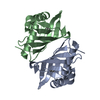 2pzvS S: Starting model for refinement |
|---|---|
| Similar structure data |


-Links
 PDBj
PDBj
- Assembly
| Deposited unit | 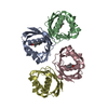
| ||||||||||||||||||||||||||||||
|---|---|---|---|---|---|---|---|---|---|---|---|---|---|---|---|---|---|---|---|---|---|---|---|---|---|---|---|---|---|---|---|
| 1 | 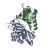
| ||||||||||||||||||||||||||||||
| 2 | 
| ||||||||||||||||||||||||||||||
| Unit cell |
| ||||||||||||||||||||||||||||||
| Noncrystallographic symmetry (NCS) | NCS domain:
NCS domain segments:
| ||||||||||||||||||||||||||||||
| Details | The biological unit is a dimer. There are 2 biological units in the asymmetric unit (chains A & B and chains C & D). |
-Components
| #1: Protein | Mass: 14471.419 Da / Num. of mol.: 4 / Mutation: Y16S,D40N Source method: isolated from a genetically manipulated source Source: (gene. exp.) Pseudomonas putida (bacteria) / Gene: ksi / Plasmid: pET21c / Production host: #2: Chemical | ChemComp-EQU /   Mass: 266.334 Da / Num. of mol.: 4 / Source method: obtained synthetically / Formula: C18H18O2 Mass: 266.334 Da / Num. of mol.: 4 / Source method: obtained synthetically / Formula: C18H18O2#3: Water | ChemComp-HOH / |  Mass: 18.015 Da / Num. of mol.: 221 / Source method: isolated from a natural source / Formula: H2O Mass: 18.015 Da / Num. of mol.: 221 / Source method: isolated from a natural source / Formula: H2O |
|---|
-Experimental details
-Experiment
| Experiment | Method: X-RAY DIFFRACTION / Number of used crystals: 1 |
|---|
- Sample preparation
| Crystal | Density Matthews: 2.58 Å3/Da / Density % sol: 52.28 % |
|---|---|
| Crystal grow | Temperature: 293 K / Method: vapor diffusion, hanging drop / pH: 7.2 Details: 0.9 M ammonium sulfate, 40 mM potassium phosphate, pH 7.2, hanging drop, temperature 293K, VAPOR DIFFUSION, HANGING DROP |
-Data collection
| Diffraction | Mean temperature: 100 K |
|---|---|
| Diffraction source | Source: SYNCHROTRON / Site: ALS  / Beamline: 8.2.2 / Wavelength: 1 Å / Beamline: 8.2.2 / Wavelength: 1 Å |
| Detector | Type: ADSC QUANTUM 315 / Detector: CCD / Date: Nov 19, 2008 |
| Radiation | Monochromator: Double crystal, Si(111) / Protocol: SINGLE WAVELENGTH / Monochromatic (M) / Laue (L): M / Scattering type: x-ray |
| Radiation wavelength | Wavelength: 1 Å / Relative weight: 1 |
| Reflection | Resolution: 1.63→43.41 Å / Num. obs: 60155 / % possible obs: 82.3 % / Biso Wilson estimate: 22.09 Å2 / Rmerge(I) obs: 0.166 / Net I/σ(I): 9.7 |
| Reflection shell | Highest resolution: 1.63 Å / Rmerge(I) obs: 0.166 / Mean I/σ(I) obs: 9.7 |
- Processing
| Software |
| |||||||||||||||||||||||||||||||||||||||||||||||||||||||||||||||||||||||||||||||||||||||||||||||||||||||||||||||||||||||||||||||||||||||||||||||||||||||||||||||||
|---|---|---|---|---|---|---|---|---|---|---|---|---|---|---|---|---|---|---|---|---|---|---|---|---|---|---|---|---|---|---|---|---|---|---|---|---|---|---|---|---|---|---|---|---|---|---|---|---|---|---|---|---|---|---|---|---|---|---|---|---|---|---|---|---|---|---|---|---|---|---|---|---|---|---|---|---|---|---|---|---|---|---|---|---|---|---|---|---|---|---|---|---|---|---|---|---|---|---|---|---|---|---|---|---|---|---|---|---|---|---|---|---|---|---|---|---|---|---|---|---|---|---|---|---|---|---|---|---|---|---|---|---|---|---|---|---|---|---|---|---|---|---|---|---|---|---|---|---|---|---|---|---|---|---|---|---|---|---|---|---|---|---|
| Refinement | Method to determine structure: MOLECULAR REPLACEMENT Starting model: PDB 2PZV Resolution: 1.632→43.408 Å / Occupancy max: 1 / Occupancy min: 0.31 / SU ML: 1.29 / Isotropic thermal model: Isotropic / σ(F): 0.09 / Phase error: 30.97 / Stereochemistry target values: ML
| |||||||||||||||||||||||||||||||||||||||||||||||||||||||||||||||||||||||||||||||||||||||||||||||||||||||||||||||||||||||||||||||||||||||||||||||||||||||||||||||||
| Solvent computation | Shrinkage radii: 0.9 Å / VDW probe radii: 1.11 Å / Solvent model: FLAT BULK SOLVENT MODEL / Bsol: 49.762 Å2 / ksol: 0.351 e/Å3 | |||||||||||||||||||||||||||||||||||||||||||||||||||||||||||||||||||||||||||||||||||||||||||||||||||||||||||||||||||||||||||||||||||||||||||||||||||||||||||||||||
| Displacement parameters | Biso mean: 50.786 Å2
| |||||||||||||||||||||||||||||||||||||||||||||||||||||||||||||||||||||||||||||||||||||||||||||||||||||||||||||||||||||||||||||||||||||||||||||||||||||||||||||||||
| Refinement step | Cycle: LAST / Resolution: 1.632→43.408 Å
| |||||||||||||||||||||||||||||||||||||||||||||||||||||||||||||||||||||||||||||||||||||||||||||||||||||||||||||||||||||||||||||||||||||||||||||||||||||||||||||||||
| Refine LS restraints |
| |||||||||||||||||||||||||||||||||||||||||||||||||||||||||||||||||||||||||||||||||||||||||||||||||||||||||||||||||||||||||||||||||||||||||||||||||||||||||||||||||
| Refine LS restraints NCS |
| |||||||||||||||||||||||||||||||||||||||||||||||||||||||||||||||||||||||||||||||||||||||||||||||||||||||||||||||||||||||||||||||||||||||||||||||||||||||||||||||||
| LS refinement shell |
| |||||||||||||||||||||||||||||||||||||||||||||||||||||||||||||||||||||||||||||||||||||||||||||||||||||||||||||||||||||||||||||||||||||||||||||||||||||||||||||||||
| Refinement TLS params. | Refine-ID: X-RAY DIFFRACTION
| |||||||||||||||||||||||||||||||||||||||||||||||||||||||||||||||||||||||||||||||||||||||||||||||||||||||||||||||||||||||||||||||||||||||||||||||||||||||||||||||||
| Refinement TLS group | Selection details: chain D |
