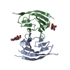
 Movie
Movie Controller

[English] 日本語
 Yorodumi
- PDB-2pzv: Crystal Structure of Ketosteroid Isomerase D40N from Pseudomonas ... -
+ Open data

- Basic information
| Entry | Database: PDB / ID: 2pzv | |||||||||
|---|---|---|---|---|---|---|---|---|---|---|
| Title | Crystal Structure of Ketosteroid Isomerase D40N from Pseudomonas Putida (pksi) with bound Phenol | |||||||||
 Components Components | Steroid Delta-isomerase | |||||||||
 Keywords Keywords | ISOMERASE / transition state analog bound | |||||||||
| Function / homology |  Function and homology information Function and homology informationsteroid Delta-isomerase / steroid Delta-isomerase activity / steroid metabolic process Similarity search - Function | |||||||||
| Biological species |  Pseudomonas putida (bacteria) Pseudomonas putida (bacteria) | |||||||||
| Method |  X-RAY DIFFRACTION / SYNCHROTRON / MOLECULAR REPLACEMENT / Resolution: 1.25 Å X-RAY DIFFRACTION / SYNCHROTRON / MOLECULAR REPLACEMENT / Resolution: 1.25 Å | |||||||||
 Authors Authors | Pybus, B. / Caaveiro, J.M.M. / Petsko, G.A. / Ringe, D. | |||||||||
 Citation Citation | Journal: PLoS Biol. / Year: 2006 Title: Testing Electrostatic complementarity in Enzyme Catalysis: Hydrogen Bonding in the Ketosteroid Isomerase Oxyanion Hole Authors: Kraut, D.A. / Sigala, P.A. / Pybus, B. / Liu, C.W. / Ringe, D. / Petsko, G.A. / Herschlag, D. | |||||||||
| History |
|
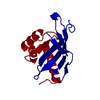
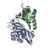
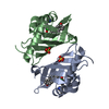
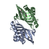
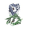
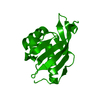
- Structure visualization
| Structure viewer | Molecule: MolmilJmol/JSmol |
|---|
- Downloads & links
-Download
| PDBx/mmCIF format | 2pzv.cif.gz | 220.5 KB | Display | PDBx/mmCIF format |
|---|---|---|---|---|
| PDB format | pdb2pzv.ent.gz | 179.2 KB | Display | PDB format |
| PDBx/mmJSON format | 2pzv.json.gz | Tree view | PDBx/mmJSON format | |
| Others |  Other downloads Other downloads |
-Validation report
| Arichive directory | https://data.pdbj.org/pub/pdb/validation_reports/pz/2pzvftp://data.pdbj.org/pub/pdb/validation_reports/pz/2pzv | HTTPS FTP |
|---|
-Related structure data
| Similar structure data |
|---|


-Links
 PDBj
PDBj

- Assembly
| Deposited unit | 
| ||||||||
|---|---|---|---|---|---|---|---|---|---|
| 1 | 
| ||||||||
| 2 | 
| ||||||||
| Unit cell |
|
-Components
| #1: Protein | Mass: 14547.515 Da / Num. of mol.: 4 / Mutation: D40N Source method: isolated from a genetically manipulated source Source: (gene. exp.) Pseudomonas putida (bacteria) / Gene: ksi / Plasmid: pKK / Species (production host): Escherichia coli / Production host: #2: Chemical | ChemComp-IPH / 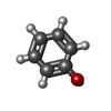  Mass: 94.111 Da / Num. of mol.: 4 / Source method: obtained synthetically / Formula: C6H6O Mass: 94.111 Da / Num. of mol.: 4 / Source method: obtained synthetically / Formula: C6H6O#3: Water | ChemComp-HOH / |  Mass: 18.015 Da / Num. of mol.: 317 / Source method: isolated from a natural source / Formula: H2O Mass: 18.015 Da / Num. of mol.: 317 / Source method: isolated from a natural source / Formula: H2O |
|---|
-Experimental details
-Experiment
| Experiment | Method: X-RAY DIFFRACTION / Number of used crystals: 1 |
|---|
- Sample preparation
| Crystal | Density Matthews: 2.06 Å3/Da / Density % sol: 40.16 % |
|---|---|
| Crystal grow | Temperature: 295 K / Method: vapor diffusion, hanging drop / pH: 7 Details: 1.4 M ammonium sulphate, 7% (v/v) 2-propanol, 40 mM potasium phosphate, VAPOR DIFFUSION, HANGING DROP, temperature 295K |
-Data collection
| Diffraction | Mean temperature: 100 K |
|---|---|
| Diffraction source | Source: SYNCHROTRON / Site: APS  / Beamline: 14-BM-C / Wavelength: 0.9 Å / Beamline: 14-BM-C / Wavelength: 0.9 Å |
| Detector | Type: ADSC QUANTUM 315 / Detector: CCD / Date: Nov 18, 2004 |
| Radiation | Protocol: SINGLE WAVELENGTH / Monochromatic (M) / Laue (L): M / Scattering type: x-ray |
| Radiation wavelength | Wavelength: 0.9 Å / Relative weight: 1 |
| Reflection | Resolution: 1.25→50 Å / Num. all: 119468 / Num. obs: 111105 / % possible obs: 93 % / Redundancy: 2.2 % / Rmerge(I) obs: 0.037 |
| Reflection shell | Resolution: 1.25→1.29 Å / Redundancy: 1.8 % / Rmerge(I) obs: 0.415 |
- Processing
| Software |
| |||||||||||||||||||||||||||||||||||||||||||||||||||||||||||||||||||||||||||||||||||||||||||||||||||||||||
|---|---|---|---|---|---|---|---|---|---|---|---|---|---|---|---|---|---|---|---|---|---|---|---|---|---|---|---|---|---|---|---|---|---|---|---|---|---|---|---|---|---|---|---|---|---|---|---|---|---|---|---|---|---|---|---|---|---|---|---|---|---|---|---|---|---|---|---|---|---|---|---|---|---|---|---|---|---|---|---|---|---|---|---|---|---|---|---|---|---|---|---|---|---|---|---|---|---|---|---|---|---|---|---|---|---|---|
| Refinement | Method to determine structure: MOLECULAR REPLACEMENT / Resolution: 1.25→22.54 Å / Cor.coef. Fo:Fc: 0.962 / Cor.coef. Fo:Fc free: 0.947 / SU B: 2.344 / SU ML: 0.044 / Isotropic thermal model: anisotropic / Cross valid method: THROUGHOUT / σ(F): 0 / ESU R: 0.067 / ESU R Free: 0.065 / Stereochemistry target values: MAXIMUM LIKELIHOOD / Details: hydrogens have been added in the riding positions
| |||||||||||||||||||||||||||||||||||||||||||||||||||||||||||||||||||||||||||||||||||||||||||||||||||||||||
| Solvent computation | Ion probe radii: 0.8 Å / Shrinkage radii: 0.8 Å / VDW probe radii: 1.4 Å / Solvent model: BABINET MODEL WITH MASK | |||||||||||||||||||||||||||||||||||||||||||||||||||||||||||||||||||||||||||||||||||||||||||||||||||||||||
| Displacement parameters | Biso mean: 26.57 Å2
| |||||||||||||||||||||||||||||||||||||||||||||||||||||||||||||||||||||||||||||||||||||||||||||||||||||||||
| Refinement step | Cycle: LAST / Resolution: 1.25→22.54 Å
| |||||||||||||||||||||||||||||||||||||||||||||||||||||||||||||||||||||||||||||||||||||||||||||||||||||||||
| Refine LS restraints |
| |||||||||||||||||||||||||||||||||||||||||||||||||||||||||||||||||||||||||||||||||||||||||||||||||||||||||
| LS refinement shell | Resolution: 1.25→1.282 Å / Total num. of bins used: 20
|
