 Movie
Movie Controller


+ Open data

- Basic information
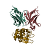
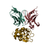
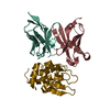
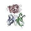
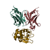
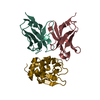
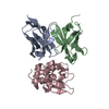
| Entry | Database: PDB / ID: 1c08 | ||||||
|---|---|---|---|---|---|---|---|
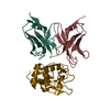
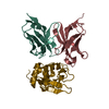
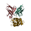
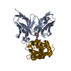
| Title | CRYSTAL STRUCTURE OF HYHEL-10 FV-HEN LYSOZYME COMPLEX | ||||||
 Components Components |
| ||||||
 Keywords Keywords | IMMUNE SYSTEM/HYDROLASE / ANTIGEN-ANTIBODY COMPLEX / IMMUNE SYSTEM-HYDROLASE COMPLEX | ||||||
| Function / homology |  Function and homology information Function and homology informationimmunoglobulin mediated immune response / immunoglobulin complex / antigen binding / Lactose synthesis / Antimicrobial peptides / Neutrophil degranulation / beta-N-acetylglucosaminidase activity / cell wall macromolecule catabolic process / lysozyme / lysozyme activity ...immunoglobulin mediated immune response / immunoglobulin complex / antigen binding / Lactose synthesis / Antimicrobial peptides / Neutrophil degranulation / beta-N-acetylglucosaminidase activity / cell wall macromolecule catabolic process / lysozyme / lysozyme activity / killing of cells of another organism / defense response to Gram-negative bacterium / adaptive immune response / defense response to bacterium / defense response to Gram-positive bacterium / immune response / endoplasmic reticulum / : / extracellular region / identical protein binding / plasma membrane / cytoplasm Similarity search - Function | ||||||
| Biological species |   | ||||||
| Method |  X-RAY DIFFRACTION / Resolution: 2.3 Å X-RAY DIFFRACTION / Resolution: 2.3 Å | ||||||
 Authors Authors | Shiroishi, M. / Kondo, H. / Matsushima, M. / Tsumoto, K. / Kumagai, I. | ||||||
 Citation Citation | Journal: J.Biol.Chem. / Year: 1999 Title: Crystal structure of anti-Hen egg white lysozyme antibody (HyHEL-10) Fv-antigen complex. Local structural changes in the protein antigen and water-mediated interactions of Fv-antigen and light ...Title: Crystal structure of anti-Hen egg white lysozyme antibody (HyHEL-10) Fv-antigen complex. Local structural changes in the protein antigen and water-mediated interactions of Fv-antigen and light chain-heavy chain interfaces. Authors: Kondo, H. / Shiroishi, M. / Matsushima, M. / Tsumoto, K. / Kumagai, I. | ||||||
| History |
| ||||||
| Remark 999 | SEQEUNCE THE SEQUENCE FOR CHAIN H IS FROM THE PRF DATABASE ACCESSION CODE 1306354A THE AUTHORS ...SEQEUNCE THE SEQUENCE FOR CHAIN H IS FROM THE PRF DATABASE ACCESSION CODE 1306354A THE AUTHORS BELIEVE THAT ALA114 IS CORRECT. |
- Structure visualization
| Structure viewer | Molecule: MolmilJmol/JSmol |
|---|
- Downloads & links
-Download
| PDBx/mmCIF format | 1c08.cif.gz | 81.5 KB | Display | PDBx/mmCIF format |
|---|---|---|---|---|
| PDB format | pdb1c08.ent.gz | 62.1 KB | Display | PDB format |
| PDBx/mmJSON format | 1c08.json.gz | Tree view | PDBx/mmJSON format | |
| Others |  Other downloads Other downloads |
-Validation report
| Arichive directory | https://data.pdbj.org/pub/pdb/validation_reports/c0/1c08ftp://data.pdbj.org/pub/pdb/validation_reports/c0/1c08 | HTTPS FTP |
|---|
-Related structure data
| Similar structure data |
|---|


-Links
 PDBj
PDBj


- Assembly
| Deposited unit | 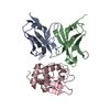
| ||||||||
|---|---|---|---|---|---|---|---|---|---|
| 1 |
| ||||||||
| Unit cell |
|
-Components
| #1: Antibody | Mass: 11623.810 Da / Num. of mol.: 1 / Fragment: VL FRAGMENT Source method: isolated from a genetically manipulated source Source: (gene. exp.)  |
|---|---|
| #2: Antibody | Mass: 12795.939 Da / Num. of mol.: 1 / Fragment: VH FRAGMENT Source method: isolated from a genetically manipulated source Source: (gene. exp.) |
| #3: Protein | Mass: 14331.160 Da / Num. of mol.: 1 / Source method: isolated from a natural source / Source: (natural) |
| #4: Water | ChemComp-HOH /  Mass: 18.015 Da / Num. of mol.: 125 / Source method: isolated from a natural source / Formula: H2O Mass: 18.015 Da / Num. of mol.: 125 / Source method: isolated from a natural source / Formula: H2O |
| Has protein modification | Y |
-Experimental details
-Experiment
| Experiment | Method: X-RAY DIFFRACTION / Number of used crystals: 1 |
|---|
- Sample preparation
| Crystal | Density Matthews: 2.49 Å3/Da / Density % sol: 50.63 % | ||||||||||||||||||||
|---|---|---|---|---|---|---|---|---|---|---|---|---|---|---|---|---|---|---|---|---|---|
| Crystal grow | Temperature: 293 K / Method: vapor diffusion, hanging drop / pH: 7.6 Details: HEPES, PEG6000, MPD, pH 7.6, VAPOR DIFFUSION, HANGING DROP, temperature 293K | ||||||||||||||||||||
| Crystal grow | *PLUS Temperature: 20 ℃ / PH range low: 7.8 / PH range high: 7.6 | ||||||||||||||||||||
| Components of the solutions | *PLUS
|
-Data collection
| Diffraction | Mean temperature: 277 K |
|---|---|
| Diffraction source | Source: ROTATING ANODE / Type: MACSCIENCE / Wavelength: 1.5418 |
| Detector | Type: MACSCIENCE / Detector: IMAGE PLATE / Date: Sep 15, 1997 |
| Radiation | Protocol: SINGLE WAVELENGTH / Monochromatic (M) / Laue (L): M / Scattering type: x-ray |
| Radiation wavelength | Wavelength: 1.5418 Å / Relative weight: 1 |
| Reflection | Resolution: 2.3→8 Å / Num. all: 146870 / Num. obs: 18301 / % possible obs: 98.5 % / Observed criterion σ(I): 0 / Redundancy: 8 % / Biso Wilson estimate: 33.2 Å2 / Rmerge(I) obs: 0.076 / Net I/σ(I): 11 |
| Reflection shell | Resolution: 2.3→2.4 Å / Redundancy: 8 % / Rmerge(I) obs: 0.265 / Num. unique all: 1771 / % possible all: 97.6 |
| Reflection shell | *PLUS % possible obs: 97.6 % |
- Processing
| Software |
| |||||||||||||||||||||||||
|---|---|---|---|---|---|---|---|---|---|---|---|---|---|---|---|---|---|---|---|---|---|---|---|---|---|---|
| Refinement | Resolution: 2.3→8 Å / σ(F): 0 / Details: Program X-PLOR by Brunger was also used.
| |||||||||||||||||||||||||
| Refinement step | Cycle: LAST / Resolution: 2.3→8 Å
| |||||||||||||||||||||||||
| Refine LS restraints |
| |||||||||||||||||||||||||
| Software | *PLUS Name: REFMAC / Classification: refinement | |||||||||||||||||||||||||
| Refinement | *PLUS Rfactor Rfree: 0.235 / Rfactor Rwork: 0.175 | |||||||||||||||||||||||||
| Solvent computation | *PLUS | |||||||||||||||||||||||||
| Displacement parameters | *PLUS |
