 Movie
Movie Controller

[English] 日本語
 Yorodumi
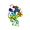
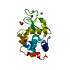
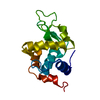
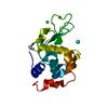
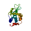
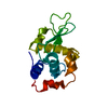
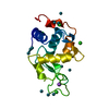
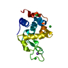
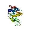
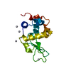
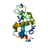
- PDB-1lz9: ANOMALOUS SIGNAL OF SOLVENT BROMINES USED FOR PHASING OF LYSOZYME -
+ Open data

- Basic information
| Entry | Database: PDB / ID: 1lz9 | ||||||
|---|---|---|---|---|---|---|---|
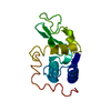
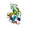
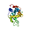
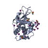
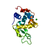
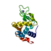
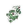
| Title | ANOMALOUS SIGNAL OF SOLVENT BROMINES USED FOR PHASING OF LYSOZYME | ||||||
 Components Components | PROTEIN (LYSOZYME) | ||||||
 Keywords Keywords | HYDROLASE / LYSOZYME / SOLVENT BROMIDES / ANOMALOUS DISPERSION / SINGLE WAVELENGTH | ||||||
| Function / homology |  Function and homology information Function and homology informationLactose synthesis / Antimicrobial peptides / Neutrophil degranulation / beta-N-acetylglucosaminidase activity / cell wall macromolecule catabolic process / lysozyme / lysozyme activity / killing of cells of another organism / defense response to Gram-negative bacterium / defense response to bacterium ...Lactose synthesis / Antimicrobial peptides / Neutrophil degranulation / beta-N-acetylglucosaminidase activity / cell wall macromolecule catabolic process / lysozyme / lysozyme activity / killing of cells of another organism / defense response to Gram-negative bacterium / defense response to bacterium / defense response to Gram-positive bacterium / endoplasmic reticulum / : / identical protein binding / cytoplasm Similarity search - Function | ||||||
| Biological species |  | ||||||
| Method |  X-RAY DIFFRACTION / SYNCHROTRON / MOLECULAR REPLACEMENT / Resolution: 1.7 Å X-RAY DIFFRACTION / SYNCHROTRON / MOLECULAR REPLACEMENT / Resolution: 1.7 Å | ||||||
 Authors Authors | Dauter, Z. / Dauter, M. | ||||||
 Citation Citation | Journal: J.Mol.Biol. / Year: 1999 Title: Anomalous signal of solvent bromides used for phasing of lysozyme. Authors: Dauter, Z. / Dauter, M. | ||||||
| History |
|
- Structure visualization
| Structure viewer | Molecule: MolmilJmol/JSmol |
|---|
- Downloads & links
-Download
| PDBx/mmCIF format | 1lz9.cif.gz | 44.6 KB | Display | PDBx/mmCIF format |
|---|---|---|---|---|
| PDB format | pdb1lz9.ent.gz | 29.9 KB | Display | PDB format |
| PDBx/mmJSON format | 1lz9.json.gz | Tree view | PDBx/mmJSON format | |
| Others |  Other downloads Other downloads |
-Validation report
| Arichive directory | https://data.pdbj.org/pub/pdb/validation_reports/lz/1lz9ftp://data.pdbj.org/pub/pdb/validation_reports/lz/1lz9 | HTTPS FTP |
|---|
-Related structure data
| Related structure data | 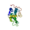 8lyzS S: Starting model for refinement |
|---|---|
| Similar structure data |


-Links
 PDBj
PDBj


- Assembly
| Deposited unit | 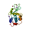
| ||||||||
|---|---|---|---|---|---|---|---|---|---|
| 1 |
| ||||||||
| Unit cell |
|
-Components
| #1: Protein | Mass: 14331.160 Da / Num. of mol.: 1 / Source method: isolated from a natural source / Details: BROMIDE AND SODIUM IONS PRESENT / Source: (natural) | ||||||
|---|---|---|---|---|---|---|---|
| #2: Chemical | ChemComp-BR /   Mass: 79.904 Da / Num. of mol.: 6 / Source method: obtained synthetically / Formula: Br Mass: 79.904 Da / Num. of mol.: 6 / Source method: obtained synthetically / Formula: Br#3: Chemical | ChemComp-NA / |   Mass: 22.990 Da / Num. of mol.: 1 / Source method: obtained synthetically / Formula: Na Mass: 22.990 Da / Num. of mol.: 1 / Source method: obtained synthetically / Formula: Na#4: Water | ChemComp-HOH / |  Mass: 18.015 Da / Num. of mol.: 197 / Source method: isolated from a natural source / Formula: H2O Mass: 18.015 Da / Num. of mol.: 197 / Source method: isolated from a natural source / Formula: H2OHas protein modification | Y | |
-Experimental details
-Experiment
| Experiment | Method: X-RAY DIFFRACTION / Number of used crystals: 1 |
|---|
- Sample preparation
| Crystal | Density Matthews: 2.05 Å3/Da / Density % sol: 40 % | ||||||||||||||||||||||||||||||||||||
|---|---|---|---|---|---|---|---|---|---|---|---|---|---|---|---|---|---|---|---|---|---|---|---|---|---|---|---|---|---|---|---|---|---|---|---|---|---|
| Crystal grow | pH: 4.6 Details: CRYSTALLIZED IN HANGING DROPS. 15 MG/ML PROTEIN SOLUTION IN 100 MM SODIUM ACETATE PH 4.6 AND 1 M SODIUM BROMIDE. | ||||||||||||||||||||||||||||||||||||
| Crystal | *PLUS | ||||||||||||||||||||||||||||||||||||
| Crystal grow | *PLUS Method: vapor diffusion, hanging drop | ||||||||||||||||||||||||||||||||||||
| Components of the solutions | *PLUS
|
-Data collection
| Diffraction | Mean temperature: 100 K | |||||||||||||||
|---|---|---|---|---|---|---|---|---|---|---|---|---|---|---|---|---|
| Diffraction source | Source: SYNCHROTRON / Site: NSLS  / Beamline: X9B / Wavelength: 0.9188, 0.9195, 0.9198, 0.9201 / Beamline: X9B / Wavelength: 0.9188, 0.9195, 0.9198, 0.9201 | |||||||||||||||
| Detector | Type: MARRESEARCH / Detector: IMAGE PLATE / Date: May 1, 1998 / Details: BENT MIRROR | |||||||||||||||
| Radiation | Monochromator: SILICON CRYSTAL / Protocol: MAD / Monochromatic (M) / Laue (L): M / Scattering type: x-ray | |||||||||||||||
| Radiation wavelength |
| |||||||||||||||
| Reflection | Resolution: 1.7→20 Å / Num. obs: 13429 / % possible obs: 99 % / Observed criterion σ(I): 0 / Redundancy: 8.5 % / Biso Wilson estimate: 16.5 Å2 / Rmerge(I) obs: 0.035 / Rsym value: 0.035 / Net I/σ(I): 50.2 | |||||||||||||||
| Reflection shell | Resolution: 1.7→1.73 Å / Redundancy: 7 % / Rmerge(I) obs: 0.182 / Mean I/σ(I) obs: 10 / Rsym value: 0.182 / % possible all: 80 |
- Processing
| Software |
| ||||||||||||||||||||||||||||||||||||||||||||||||||||||||||||||||||||||||||||||||||||
|---|---|---|---|---|---|---|---|---|---|---|---|---|---|---|---|---|---|---|---|---|---|---|---|---|---|---|---|---|---|---|---|---|---|---|---|---|---|---|---|---|---|---|---|---|---|---|---|---|---|---|---|---|---|---|---|---|---|---|---|---|---|---|---|---|---|---|---|---|---|---|---|---|---|---|---|---|---|---|---|---|---|---|---|---|---|
| Refinement | Method to determine structure: MOLECULAR REPLACEMENT Starting model: PDB ENTRY 8LYZ Resolution: 1.7→20 Å / SU B: 2.7 / SU ML: 0.094 / Cross valid method: FREE R / σ(F): 0 / ESU R: 0.152 / ESU R Free: 0.162
| ||||||||||||||||||||||||||||||||||||||||||||||||||||||||||||||||||||||||||||||||||||
| Displacement parameters | Biso mean: 17.3 Å2
| ||||||||||||||||||||||||||||||||||||||||||||||||||||||||||||||||||||||||||||||||||||
| Refinement step | Cycle: LAST / Resolution: 1.7→20 Å
| ||||||||||||||||||||||||||||||||||||||||||||||||||||||||||||||||||||||||||||||||||||
| Refine LS restraints |
| ||||||||||||||||||||||||||||||||||||||||||||||||||||||||||||||||||||||||||||||||||||
| Software | *PLUS Name: REFMAC / Classification: refinement | ||||||||||||||||||||||||||||||||||||||||||||||||||||||||||||||||||||||||||||||||||||
| Refinement | *PLUS Highest resolution: 1.7 Å | ||||||||||||||||||||||||||||||||||||||||||||||||||||||||||||||||||||||||||||||||||||
| Solvent computation | *PLUS | ||||||||||||||||||||||||||||||||||||||||||||||||||||||||||||||||||||||||||||||||||||
| Displacement parameters | *PLUS |
