 Movie
Movie Controller

[English] 日本語
 Yorodumi
- PDB-1bhz: LOW TEMPERATURE MIDDLE RESOLUTION STRUCTURE OF HEN EGG WHITE LYSO... -
+ Open data

- Basic information
| Entry | Database: PDB / ID: 1bhz | ||||||
|---|---|---|---|---|---|---|---|
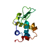
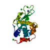
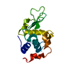
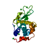
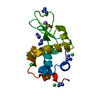
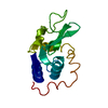
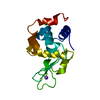
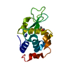
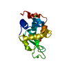
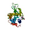
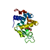
| Title | LOW TEMPERATURE MIDDLE RESOLUTION STRUCTURE OF HEN EGG WHITE LYSOZYME FROM MASC DATA | ||||||
 Components Components | LYSOZYME | ||||||
 Keywords Keywords | HYDROLASE / O-GLYCOSYL / MASC / MULTIWAVELENGTH ANOMALOUS SOLVENT CONTRAST | ||||||
| Function / homology |  Function and homology information Function and homology informationLactose synthesis / Antimicrobial peptides / Neutrophil degranulation / beta-N-acetylglucosaminidase activity / cell wall macromolecule catabolic process / lysozyme / lysozyme activity / killing of cells of another organism / defense response to Gram-negative bacterium / defense response to bacterium ...Lactose synthesis / Antimicrobial peptides / Neutrophil degranulation / beta-N-acetylglucosaminidase activity / cell wall macromolecule catabolic process / lysozyme / lysozyme activity / killing of cells of another organism / defense response to Gram-negative bacterium / defense response to bacterium / defense response to Gram-positive bacterium / endoplasmic reticulum / : / identical protein binding / cytoplasm Similarity search - Function | ||||||
| Biological species |  | ||||||
| Method |  X-RAY DIFFRACTION / SYNCHROTRON / MOLECULAR REPLACEMENT, RIGID BODY REFINEMENT / Resolution: 3.9 Å X-RAY DIFFRACTION / SYNCHROTRON / MOLECULAR REPLACEMENT, RIGID BODY REFINEMENT / Resolution: 3.9 Å | ||||||
 Authors Authors | Ramin, M. / Shepard, W. / Fourme, R. / Kahn, R. | ||||||
 Citation Citation | Journal: Acta Crystallogr.,Sect.D / Year: 1999 Title: Multiwavelength anomalous solvent contrast (MASC): derivation of envelope structure-factor amplitudes and comparison with model values. Authors: Ramin, M. / Shepard, W. / Fourme, R. / Kahn, R. #1: Journal: Nature / Year: 1965Title: Structure of Hen Egg-White Lysozyme. A Three-Dimensional Fourier Synthesis at 2 Angstrom Resolution Authors: Blake, C.C. / Koenig, D.F. / Mair, G.A. / North, A.C. / Phillips, D.C. / Sarma, V.R. | ||||||
| History |
|
- Structure visualization
| Structure viewer | Molecule: MolmilJmol/JSmol |
|---|
- Downloads & links
-Download
| PDBx/mmCIF format | 1bhz.cif.gz | 34.2 KB | Display | PDBx/mmCIF format |
|---|---|---|---|---|
| PDB format | pdb1bhz.ent.gz | 22.3 KB | Display | PDB format |
| PDBx/mmJSON format | 1bhz.json.gz | Tree view | PDBx/mmJSON format | |
| Others |  Other downloads Other downloads |
-Validation report
| Arichive directory | https://data.pdbj.org/pub/pdb/validation_reports/bh/1bhzftp://data.pdbj.org/pub/pdb/validation_reports/bh/1bhz | HTTPS FTP |
|---|
-Related structure data
| Related structure data | 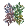 1bhwC  1bhyC 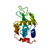 1lseS S: Starting model for refinement C: citing same article ( |
|---|---|
| Similar structure data |


-Links
 PDBj
PDBj


- Assembly
| Deposited unit | 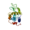
| ||||||||
|---|---|---|---|---|---|---|---|---|---|
| 1 |
| ||||||||
| Unit cell |
|
-Components
| #1: Protein | Mass: 14331.160 Da / Num. of mol.: 1 / Source method: isolated from a natural source / Source: (natural) |
|---|---|
| Has protein modification | Y |
-Experimental details
-Experiment
| Experiment | Method: X-RAY DIFFRACTION / Number of used crystals: 1 |
|---|
- Sample preparation
| Crystal | Density Matthews: 1.9 Å3/Da / Density % sol: 30.3 % |
|---|---|
| Crystal grow | Details: PROTEIN WAS CRYSTALLIZED FROM SODIUM CHLORIDE SOLUTIONS |
-Data collection
| Diffraction | Mean temperature: 124 K |
|---|---|
| Diffraction source | Source: SYNCHROTRON / Site: LURE  / Beamline: DW21B / Wavelength: 1.39 / Beamline: DW21B / Wavelength: 1.39 |
| Detector | Type: MARRESEARCH / Detector: IMAGE PLATE / Date: Nov 1, 1996 / Details: MIRRORS |
| Radiation | Monochromator: SI(111) / Monochromatic (M) / Laue (L): M / Scattering type: x-ray |
| Radiation wavelength | Wavelength: 1.39 Å / Relative weight: 1 |
| Reflection | Resolution: 3.91→33.5 Å / Num. obs: 1092 / % possible obs: 84.7 % / Observed criterion σ(I): 3 / Redundancy: 9.4 % / Rmerge(I) obs: 0.028 / Rsym value: 0.028 / Net I/σ(I): 18.3 |
| Reflection shell | Resolution: 3.91→4 Å / Redundancy: 2 % / Rmerge(I) obs: 0.028 / Mean I/σ(I) obs: 46.7 / Rsym value: 0.028 / % possible all: 78 |
| Reflection | *PLUS Num. measured all: 10257 |
- Processing
| Software |
| ||||||||||||||||||||||||
|---|---|---|---|---|---|---|---|---|---|---|---|---|---|---|---|---|---|---|---|---|---|---|---|---|---|
| Refinement | Method to determine structure: MOLECULAR REPLACEMENT, RIGID BODY REFINEMENT Starting model: 1LSE Resolution: 3.9→8 Å / Cross valid method: THROUGHOUT
| ||||||||||||||||||||||||
| Refinement step | Cycle: LAST / Resolution: 3.9→8 Å
|
