 Movie
Movie Controller

[English] 日本語
 Yorodumi
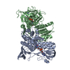
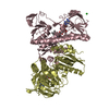
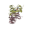
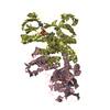
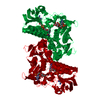
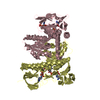
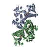
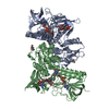
- PDB-1bhy: LOW TEMPERATURE MIDDLE RESOLUTION STRUCTURE OF P64K FROM MASC DATA -
+ Open data

- Basic information
| Entry | Database: PDB / ID: 1bhy | ||||||
|---|---|---|---|---|---|---|---|
| Title | LOW TEMPERATURE MIDDLE RESOLUTION STRUCTURE OF P64K FROM MASC DATA | ||||||
 Components Components | P64K | ||||||
 Keywords Keywords | OUTER MEMBRANE PROTEIN / MASC / MULTIWAVELENGTH ANOMALOUS SOLVENT CONTRAST | ||||||
| Function / homology |  Function and homology information Function and homology informationdihydrolipoyl dehydrogenase / dihydrolipoyl dehydrogenase (NADH) activity / 2-oxoglutarate metabolic process / flavin adenine dinucleotide binding Similarity search - Function | ||||||
| Biological species |  Neisseria meningitidis (bacteria) Neisseria meningitidis (bacteria) | ||||||
| Method |  X-RAY DIFFRACTION / SYNCHROTRON / MOLECULAR REPLACEMENT, RIGID BODY REFINEMENT / Resolution: 4.18 Å X-RAY DIFFRACTION / SYNCHROTRON / MOLECULAR REPLACEMENT, RIGID BODY REFINEMENT / Resolution: 4.18 Å | ||||||
 Authors Authors | Ramin, M. / Shepard, W. / Fourme, R. / Kahn, R. | ||||||
 Citation Citation | Journal: Acta Crystallogr.,Sect.D / Year: 1999 Title: Multiwavelength anomalous solvent contrast (MASC): derivation of envelope structure-factor amplitudes and comparison with model values. Authors: Ramin, M. / Shepard, W. / Fourme, R. / Kahn, R. #1: Journal: J.Mol.Biol. / Year: 1997Title: Molecular Structure of the Lipoamide Dehydrogenase Domain of a Surface Antigen from Neisseria Meningitidis Authors: Li De La Sierra, I. / Pernot, L. / Prange, T. / Saludjian, P. / Schiltz, M. / Fourme, R. / Padron, G. | ||||||
| History |
|
- Structure visualization
| Structure viewer | Molecule: MolmilJmol/JSmol |
|---|
- Downloads & links
-Download
| PDBx/mmCIF format | 1bhy.cif.gz | 94.5 KB | Display | PDBx/mmCIF format |
|---|---|---|---|---|
| PDB format | pdb1bhy.ent.gz | 67.9 KB | Display | PDB format |
| PDBx/mmJSON format | 1bhy.json.gz | Tree view | PDBx/mmJSON format | |
| Others |  Other downloads Other downloads |
-Validation report
| Arichive directory | https://data.pdbj.org/pub/pdb/validation_reports/bh/1bhyftp://data.pdbj.org/pub/pdb/validation_reports/bh/1bhy | HTTPS FTP |
|---|
-Related structure data
| Related structure data | 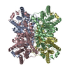 1bhwC 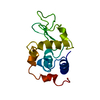 1bhzC  1ojtS S: Starting model for refinement C: citing same article ( |
|---|---|
| Similar structure data |


-Links
 PDBj
PDBj


- Assembly
| Deposited unit | 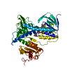
| ||||||||
|---|---|---|---|---|---|---|---|---|---|
| 1 | 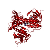
| ||||||||
| Unit cell |
|
-Components
| #1: Protein | Mass: 50797.992 Da / Num. of mol.: 1 / Source method: isolated from a natural source / Source: (natural) Neisseria meningitidis (bacteria) / References: UniProt: Q51225 |
|---|---|
| #2: Chemical | ChemComp-FAD /   Mass: 785.550 Da / Num. of mol.: 1 / Source method: obtained synthetically / Formula: C27H33N9O15P2 / Comment: FAD*YM Mass: 785.550 Da / Num. of mol.: 1 / Source method: obtained synthetically / Formula: C27H33N9O15P2 / Comment: FAD*YM |
| Has protein modification | Y |
-Experimental details
-Experiment
| Experiment | Method: X-RAY DIFFRACTION / Number of used crystals: 1 |
|---|
- Sample preparation
| Crystal | Density Matthews: 3.6 Å3/Da / Density % sol: 60.5 % | ||||||||||||||||||||||||||||||||||||||||||
|---|---|---|---|---|---|---|---|---|---|---|---|---|---|---|---|---|---|---|---|---|---|---|---|---|---|---|---|---|---|---|---|---|---|---|---|---|---|---|---|---|---|---|---|
| Crystal grow | Details: PROTEIN WAS CRYSTALLIZED FROM AMMONIUM SULPHATE | ||||||||||||||||||||||||||||||||||||||||||
| Crystal | *PLUS | ||||||||||||||||||||||||||||||||||||||||||
| Crystal grow | *PLUS pH: 7.2 / Method: vapor diffusion, hanging dropDetails: Li De La Sierra, I., (1994) J.Mol.Biol., 235, 1154. | ||||||||||||||||||||||||||||||||||||||||||
| Components of the solutions | *PLUS
|
-Data collection
| Diffraction | Mean temperature: 124 K |
|---|---|
| Diffraction source | Source: SYNCHROTRON / Site: ESRF  / Type: ESRF / Wavelength: 0.99 / Type: ESRF / Wavelength: 0.99 |
| Detector | Type: MARRESEARCH / Detector: IMAGE PLATE / Date: Jun 1, 1995 / Details: MIRRORS |
| Radiation | Monochromator: SI(111) / Monochromatic (M) / Laue (L): M / Scattering type: x-ray |
| Radiation wavelength | Wavelength: 0.99 Å / Relative weight: 1 |
| Reflection | Resolution: 4.18→100 Å / Num. obs: 6008 / % possible obs: 98.2 % / Observed criterion σ(I): 3 / Redundancy: 7.4 % / Rmerge(I) obs: 0.055 / Rsym value: 0.055 / Net I/σ(I): 5.2 |
| Reflection shell | Resolution: 4.18→4.29 Å / Redundancy: 4.2 % / Rmerge(I) obs: 0.06 / Mean I/σ(I) obs: 10.7 / Rsym value: 0.06 / % possible all: 78 |
| Reflection | *PLUS Num. measured all: 44222 |
- Processing
| Software |
| ||||||||||||||||||||||||
|---|---|---|---|---|---|---|---|---|---|---|---|---|---|---|---|---|---|---|---|---|---|---|---|---|---|
| Refinement | Method to determine structure: MOLECULAR REPLACEMENT, RIGID BODY REFINEMENT Starting model: 1OJT Resolution: 4.18→8 Å / Cross valid method: THROUGHOUT
| ||||||||||||||||||||||||
| Refinement step | Cycle: LAST / Resolution: 4.18→8 Å
|
