 Movie
Movie Controller


+ Open data

- Basic information
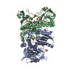
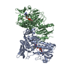
| Entry | Database: PDB / ID: 1jeh | ||||||
|---|---|---|---|---|---|---|---|
| Title | CRYSTAL STRUCTURE OF YEAST E3, LIPOAMIDE DEHYDROGENASE | ||||||
 Components Components | DIHYDROLIPOAMIDE DEHYDROGENASE | ||||||
 Keywords Keywords | OXIDOREDUCTASE / 2-oxoglutarate dehydrogenase complex / pyruvate dehydrogenase complex | ||||||
| Function / homology |  Function and homology information Function and homology informationGlycine degradation / OGDH complex synthesizes succinyl-CoA from 2-OG / PDH complex synthesizes acetyl-CoA from PYR / glycine catabolic process / glycine dehydrogenase (decarboxylating) activity / oxoglutarate dehydrogenase (succinyl-transferring) activity / glycine cleavage complex / dihydrolipoyl dehydrogenase / dihydrolipoyl dehydrogenase (NADH) activity / L-valine catabolic process ...Glycine degradation / OGDH complex synthesizes succinyl-CoA from 2-OG / PDH complex synthesizes acetyl-CoA from PYR / glycine catabolic process / glycine dehydrogenase (decarboxylating) activity / oxoglutarate dehydrogenase (succinyl-transferring) activity / glycine cleavage complex / dihydrolipoyl dehydrogenase / dihydrolipoyl dehydrogenase (NADH) activity / L-valine catabolic process / L-isoleucine catabolic process / hydrogen peroxide metabolic process / L-leucine catabolic process / oxoglutarate dehydrogenase complex / pyruvate decarboxylation to acetyl-CoA / L-serine biosynthetic process / pyruvate dehydrogenase complex / 2-oxoglutarate metabolic process / cellular respiration / mitochondrial nucleoid / tricarboxylic acid cycle / flavin adenine dinucleotide binding / mitochondrion Similarity search - Function | ||||||
| Biological species |  | ||||||
| Method |  X-RAY DIFFRACTION / SYNCHROTRON / MOLECULAR REPLACEMENT / Resolution: 2.4 Å X-RAY DIFFRACTION / SYNCHROTRON / MOLECULAR REPLACEMENT / Resolution: 2.4 Å | ||||||
 Authors Authors | Toyoda, T. / Suzuki, K. / Sekigushi, T. / Reed, J. / Takenaka, A. | ||||||
 Citation Citation | Journal: J.Biochem. / Year: 1998 Title: Crystal structure of eucaryotic E3, lipoamide dehydrogenase from yeast. Authors: Toyoda, T. / Suzuki, K. / Sekiguchi, T. / Reed, L.J. / Takenaka, A. | ||||||
| History |
|
- Structure visualization
| Structure viewer | Molecule: MolmilJmol/JSmol |
|---|
- Downloads & links
-Download
| PDBx/mmCIF format | 1jeh.cif.gz | 192.4 KB | Display | PDBx/mmCIF format |
|---|---|---|---|---|
| PDB format | pdb1jeh.ent.gz | 153.6 KB | Display | PDB format |
| PDBx/mmJSON format | 1jeh.json.gz | Tree view | PDBx/mmJSON format | |
| Others |  Other downloads Other downloads |
-Validation report
| Arichive directory | https://data.pdbj.org/pub/pdb/validation_reports/je/1jehftp://data.pdbj.org/pub/pdb/validation_reports/je/1jeh | HTTPS FTP |
|---|
-Related structure data
| Related structure data |  3grsS S: Starting model for refinement |
|---|---|
| Similar structure data |


-Links
 PDBj
PDBj


- Assembly
| Deposited unit | 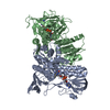
| ||||||||
|---|---|---|---|---|---|---|---|---|---|
| 1 |
| ||||||||
| Unit cell |
|
-Components
| #1: Protein | Mass: 51623.992 Da / Num. of mol.: 2 / Source method: isolated from a natural source / Source: (natural) #2: Chemical |   Mass: 785.550 Da / Num. of mol.: 2 / Source method: obtained synthetically / Formula: C27H33N9O15P2 / Comment: FAD*YM Mass: 785.550 Da / Num. of mol.: 2 / Source method: obtained synthetically / Formula: C27H33N9O15P2 / Comment: FAD*YM#3: Water | ChemComp-HOH / |  Mass: 18.015 Da / Num. of mol.: 73 / Source method: isolated from a natural source / Formula: H2O Mass: 18.015 Da / Num. of mol.: 73 / Source method: isolated from a natural source / Formula: H2OHas protein modification | Y | |
|---|
-Experimental details
-Experiment
| Experiment | Method: X-RAY DIFFRACTION / Number of used crystals: 1 |
|---|
- Sample preparation
| Crystal | Density Matthews: 2.5 Å3/Da / Density % sol: 50.8 % | ||||||||||||||||||||||||
|---|---|---|---|---|---|---|---|---|---|---|---|---|---|---|---|---|---|---|---|---|---|---|---|---|---|
| Crystal grow | Temperature: 298 K / Method: microdialysis / pH: 7 / Details: PEG6000, pH 7.00, MICRODIALYSIS, temperature 298K | ||||||||||||||||||||||||
| Crystal grow | *PLUS Details: Toyoda, T., (1997) J.Biochem.(Tokyo), 121, 1. | ||||||||||||||||||||||||
| Components of the solutions | *PLUS
|
-Data collection
| Diffraction | Mean temperature: 298 K |
|---|---|
| Diffraction source | Source: SYNCHROTRON / Site: Photon Factory  / Beamline: BL-18B / Wavelength: 1 / Wavelength: 1 Å / Beamline: BL-18B / Wavelength: 1 / Wavelength: 1 Å |
| Detector | Type: FUJI / Detector: IMAGE PLATE |
| Radiation | Monochromator: GRAPHITE / Protocol: SINGLE WAVELENGTH / Monochromatic (M) / Laue (L): M / Scattering type: x-ray |
| Radiation wavelength | Wavelength: 1 Å / Relative weight: 1 |
| Reflection | Resolution: 2.4→100 Å / Num. obs: 33372 / % possible obs: 80.2 % / Observed criterion σ(F): 2 / Observed criterion σ(I): 2 / Redundancy: 2.9 % / Rmerge(I) obs: 0.067 |
| Reflection shell | Resolution: 2.4→2.68 Å / % possible all: 42.7 |
| Reflection | *PLUS Highest resolution: 2.4 Å / Lowest resolution: 100 Å / Redundancy: 2.9 % / Num. measured all: 97677 |
- Processing
| Software |
| ||||||||||||||||||||||||||||||||||||||||||||||||||||||||||||
|---|---|---|---|---|---|---|---|---|---|---|---|---|---|---|---|---|---|---|---|---|---|---|---|---|---|---|---|---|---|---|---|---|---|---|---|---|---|---|---|---|---|---|---|---|---|---|---|---|---|---|---|---|---|---|---|---|---|---|---|---|---|
| Refinement | Method to determine structure: MOLECULAR REPLACEMENT Starting model: PDB ENTRY 3GRS Resolution: 2.4→10 Å / σ(F): 4
| ||||||||||||||||||||||||||||||||||||||||||||||||||||||||||||
| Displacement parameters | Biso mean: 35.7 Å2 | ||||||||||||||||||||||||||||||||||||||||||||||||||||||||||||
| Refine analyze | Luzzati coordinate error obs: 0.35 Å | ||||||||||||||||||||||||||||||||||||||||||||||||||||||||||||
| Refinement step | Cycle: LAST / Resolution: 2.4→10 Å
| ||||||||||||||||||||||||||||||||||||||||||||||||||||||||||||
| Refine LS restraints |
| ||||||||||||||||||||||||||||||||||||||||||||||||||||||||||||
| LS refinement shell | Resolution: 2.4→2.51 Å / Total num. of bins used: 8 /
| ||||||||||||||||||||||||||||||||||||||||||||||||||||||||||||
| Software | *PLUS Name: X-PLOR / Version: 3.1 / Classification: refinement | ||||||||||||||||||||||||||||||||||||||||||||||||||||||||||||
| Refinement | *PLUS % reflection Rfree: 10 % / Rfactor Rfree: 0.26 | ||||||||||||||||||||||||||||||||||||||||||||||||||||||||||||
| Solvent computation | *PLUS | ||||||||||||||||||||||||||||||||||||||||||||||||||||||||||||
| Displacement parameters | *PLUS Biso mean: 35.7 Å2 | ||||||||||||||||||||||||||||||||||||||||||||||||||||||||||||
| Refine LS restraints | *PLUS Type: x_improper_angle_d / Dev ideal: 0.763 | ||||||||||||||||||||||||||||||||||||||||||||||||||||||||||||
| LS refinement shell | *PLUS Rfactor Rfree: 0.303 / % reflection Rfree: 10 % / Rfactor Rwork: 0.286 |
