response to hypochlorite / oxidoreductase activity, acting on a sulfur group of donors, NAD(P) as acceptor / cupric reductase (NADH) activity / NAD(P)H dehydrogenase (quinone) activity / oxidoreductase activity, acting on NAD(P)H / flavin adenine dinucleotide binding / protein homodimerization activity Similarity search - Function
: / Pyridine nucleotide-disulphide oxidoreductase / Pyridine nucleotide-disulphide oxidoreductase, class I / Pyridine nucleotide-disulphide oxidoreductase, class I, active site / Pyridine nucleotide-disulphide oxidoreductases class-I active site. / Pyridine nucleotide-disulphide oxidoreductase, dimerisation domain / Pyridine nucleotide-disulphide oxidoreductase, dimerisation domain / FAD/NAD-linked reductase, dimerisation domain superfamily / FAD/NAD(P)-binding domain / Pyridine nucleotide-disulphide oxidoreductase / FAD/NAD(P)-binding domain superfamily Similarity search - Domain/homology
Method to determine structure: MOLECULAR REPLACEMENT Starting model: sequence based de novo model (Galaxy Web server) Resolution: 2.9→40.37 Å / SU ML: 0.4122 / Cross valid method: FREE R-VALUE / σ(F): 1.58 / Phase error: 27.3977
Rfactor
Num. reflection
% reflection
Rfree
0.2793
2010
3.72 %
Rwork
0.233
-
-
obs
0.2348
54069
96.43 %
Solvent computation
Shrinkage radii: 0.9 Å / VDW probe radii: 1.11 Å
Displacement parameters
Biso mean: 39.43 Å2
Refinement step
Cycle: LAST / Resolution: 2.9→40.37 Å
Protein
Nucleic acid
Ligand
Solvent
Total
Num. atoms
13495
0
215
26
13736
Refine LS restraints
Refine-ID
Type
Dev ideal
Number
X-RAY DIFFRACTION
f_bond_d
0.0026
13994
X-RAY DIFFRACTION
f_angle_d
0.5293
19034
X-RAY DIFFRACTION
f_chiral_restr
0.043
2204
X-RAY DIFFRACTION
f_plane_restr
0.0031
2463
X-RAY DIFFRACTION
f_dihedral_angle_d
8.1982
9900
LS refinement shell
Resolution (Å)
Rfactor Rfree
Num. reflection Rfree
Rfactor Rwork
Num. reflection Rwork
Refine-ID
% reflection obs (%)
2.9-2.94
0.3075
100
0.2595
2710
X-RAY DIFFRACTION
70.43
2.94-3.02
0.3577
148
0.2658
3611
X-RAY DIFFRACTION
93.41
3.02-3.11
0.3145
152
0.2465
3660
X-RAY DIFFRACTION
96.09
3.11-3.21
0.28
136
0.2448
3736
X-RAY DIFFRACTION
97.31
3.21-3.32
0.2914
142
0.2528
3805
X-RAY DIFFRACTION
98.36
3.32-3.46
0.3004
149
0.2533
3780
X-RAY DIFFRACTION
98.67
3.46-3.61
0.3178
141
0.2428
3851
X-RAY DIFFRACTION
99.2
3.61-3.8
0.2798
153
0.2293
3803
X-RAY DIFFRACTION
99
3.8-4.04
0.2828
139
0.2175
3848
X-RAY DIFFRACTION
99.55
4.04-4.35
0.2501
142
0.2153
3820
X-RAY DIFFRACTION
99.77
4.35-4.79
0.2751
157
0.2091
3865
X-RAY DIFFRACTION
99.73
4.79-5.48
0.2523
151
0.2236
3829
X-RAY DIFFRACTION
99.62
5.48-6.9
0.2789
146
0.25
3865
X-RAY DIFFRACTION
99.9
6.9-40.37
0.2464
154
0.2269
3876
X-RAY DIFFRACTION
98.85
+
About Yorodumi
-
News
-
Feb 9, 2022. New format data for meta-information of EMDB entries
New format data for meta-information of EMDB entries
Version 3 of the EMDB header file is now the official format.
The previous official version 1.9 will be removed from the archive.
In the structure databanks used in Yorodumi, some data are registered as the other names, "COVID-19 virus" and "2019-nCoV". Here are the details of the virus and the list of structure data.
Jan 31, 2019. EMDB accession codes are about to change! (news from PDBe EMDB page)
EMDB accession codes are about to change! (news from PDBe EMDB page)
The allocation of 4 digits for EMDB accession codes will soon come to an end. Whilst these codes will remain in use, new EMDB accession codes will include an additional digit and will expand incrementally as the available range of codes is exhausted. The current 4-digit format prefixed with “EMD-” (i.e. EMD-XXXX) will advance to a 5-digit format (i.e. EMD-XXXXX), and so on. It is currently estimated that the 4-digit codes will be depleted around Spring 2019, at which point the 5-digit format will come into force.
The EM Navigator/Yorodumi systems omit the EMD- prefix.
Related info.:Q: What is EMD? / ID/Accession-code notation in Yorodumi/EM Navigator
Yorodumi is a browser for structure data from EMDB, PDB, SASBDB, etc.
This page is also the successor to EM Navigator detail page, and also detail information page/front-end page for Omokage search.
The word "yorodu" (or yorozu) is an old Japanese word meaning "ten thousand". "mi" (miru) is to see.
Related info.:EMDB / PDB / SASBDB / Comparison of 3 databanks / Yorodumi Search / Aug 31, 2016. New EM Navigator & Yorodumi / Yorodumi Papers / Jmol/JSmol / Function and homology information / Changes in new EM Navigator and Yorodumi
 Movie
Movie Controller

 Yorodumi
 Open data
 Basic information
 Components
 Keywords
 Function and homology information
 X-RAY DIFFRACTION /
  Authors
 Citation
 Structure visualization
 Downloads & links
 Other downloads


 PDBj
PDBj


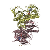
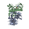
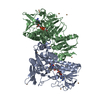
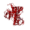
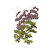
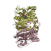
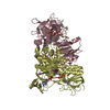
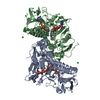
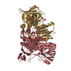
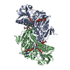
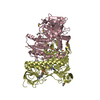
 Assembly
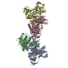
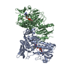
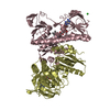

 Mass: 785.550 Da / Num. of mol.: 4 / Source method: isolated from a natural source / Formula: C27H33N9O15P2 / Feature type: SUBJECT OF INVESTIGATION / Comment: FAD*YM
 Mass: 35.453 Da / Num. of mol.: 3 / Source method: obtained synthetically / Formula: Cl
 Mass: 18.015 Da / Num. of mol.: 26 / Source method: isolated from a natural source / Formula: H2O
 Sample preparation
 / Beamline: 5C (4A) / Wavelength: 0.9796 Å
 Processing