 Movie
Movie Controller

[English] 日本語
 Yorodumi
- PDB-1dxl: Dihydrolipoamide dehydrogenase of glycine decarboxylase from Pisu... -
+ Open data

- Basic information
| Entry | Database: PDB / ID: 1dxl | ||||||
|---|---|---|---|---|---|---|---|
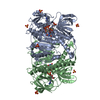
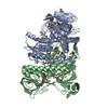
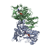
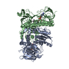
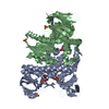
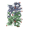
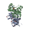
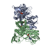
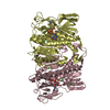
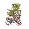
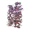
| Title | Dihydrolipoamide dehydrogenase of glycine decarboxylase from Pisum Sativum | ||||||
 Components Components | DIHYDROLIPOAMIDE DEHYDROGENASE | ||||||
 Keywords Keywords | OXIDOREDUCTASE / DIHYDROLIPOAMIDE DEHYDROGENASE / MULTIENZYME COMPLEX PROTEIN / PYRUVATE DEHYDROGENASE COMPLEX / GLYCINE DECARBOXYLASE COMPLEX / FLAVOPROTEIN | ||||||
| Function / homology |  Function and homology information Function and homology informationglycine decarboxylation via glycine cleavage system / glycine cleavage complex / dihydrolipoyl dehydrogenase / dihydrolipoyl dehydrogenase (NADH) activity / oxoglutarate dehydrogenase complex / 2-oxoglutarate metabolic process / flavin adenine dinucleotide binding / mitochondrial matrix / mitochondrion Similarity search - Function | ||||||
| Biological species |  PISUM SATIVUM (garden pea) PISUM SATIVUM (garden pea) | ||||||
| Method |  X-RAY DIFFRACTION / SYNCHROTRON / MOLECULAR REPLACEMENT / Resolution: 3.15 Å X-RAY DIFFRACTION / SYNCHROTRON / MOLECULAR REPLACEMENT / Resolution: 3.15 Å | ||||||
 Authors Authors | Faure, M. / Cohen-Addad, C. / Bourguignon, J. / Macherel, D. / Neuburger, M. / Douce, R. | ||||||
 Citation Citation | Journal: Eur.J.Biochem. / Year: 2000 Title: Interaction between the Lipoamide-Containing H-Protein and the Lipoamide Dehydrogenase (L-Protein) of the Glycine Decarboxylase Multienzyme System. 2. Crystal Structure of H- and L-Proteins Authors: Faure, M. / Bourguignon, J. / Neuburger, M. / Macherel, D. / Sieker, L. / Ober, R. / Kahn, R. / Cohen-Addad, C. / Douce, R. #1: Journal: Biochem.J. / Year: 1996 Title: Glycine Decarboxylase and Pyruvate Dehydrogenase Complexes Share the Same Dihydrolipoamide Dehydrogenase in Pea Leaf Mitochondria: Evidence from Mass Spectrometry and Primary-Structure Analysis Authors: Bourguignon, J. / Merand, V. / Rawsthorne, S. / Forest, E. / Douce, R. | ||||||
| History |
|
- Structure visualization
| Structure viewer | Molecule: MolmilJmol/JSmol |
|---|
- Downloads & links
-Download
| PDBx/mmCIF format | 1dxl.cif.gz | 331.8 KB | Display | PDBx/mmCIF format |
|---|---|---|---|---|
| PDB format | pdb1dxl.ent.gz | 273.7 KB | Display | PDB format |
| PDBx/mmJSON format | 1dxl.json.gz | Tree view | PDBx/mmJSON format | |
| Others |  Other downloads Other downloads |
-Validation report
| Arichive directory | https://data.pdbj.org/pub/pdb/validation_reports/dx/1dxlftp://data.pdbj.org/pub/pdb/validation_reports/dx/1dxl | HTTPS FTP |
|---|
-Related structure data
| Related structure data |  1dxmC  3ladS S: Starting model for refinement C: citing same article ( |
|---|---|
| Similar structure data |


-Links
 PDBj
PDBj


- Assembly
| Deposited unit | 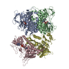
| ||||||||||||||||
|---|---|---|---|---|---|---|---|---|---|---|---|---|---|---|---|---|---|
| 1 | 
| ||||||||||||||||
| 2 | 
| ||||||||||||||||
| Unit cell |
| ||||||||||||||||
| Noncrystallographic symmetry (NCS) | NCS oper:
| ||||||||||||||||
| Details | BIOLOGICAL_UNIT: HOMODIMERTHE ASYMETRIC UNIT CONTAINS 2 DIMERS (AB AND CD) |
-Components
| #1: Protein | Mass: 49808.961 Da / Num. of mol.: 4 Source method: isolated from a genetically manipulated source Source: (gene. exp.) PISUM SATIVUM (garden pea) / Tissue: LEAF / Cellular location: MITOCHONDRIA / Organelle: MITOCHONDRIA / Plasmid: PET3A / Cellular location (production host): INCLUSION BODIES / Production host:  #2: Chemical | ChemComp-FAD /   Mass: 785.550 Da / Num. of mol.: 4 / Source method: obtained synthetically / Formula: C27H33N9O15P2 / Comment: FAD*YM Mass: 785.550 Da / Num. of mol.: 4 / Source method: obtained synthetically / Formula: C27H33N9O15P2 / Comment: FAD*YM#3: Water | ChemComp-HOH / |  Mass: 18.015 Da / Num. of mol.: 104 / Source method: isolated from a natural source / Formula: H2O Mass: 18.015 Da / Num. of mol.: 104 / Source method: isolated from a natural source / Formula: H2OHas protein modification | Y | Sequence details | THE SWISSPROT ENTRY P31023 GIVES THE SEQUENCE FROM BOURGUIGNON J., MACHEREL D., NEUBURGER M., DOUCE ...THE SWISSPROT ENTRY P31023 GIVES THE SEQUENCE FROM BOURGUIGNO | |
|---|
-Experimental details
-Experiment
| Experiment | Method: X-RAY DIFFRACTION / Number of used crystals: 1 |
|---|
- Sample preparation
| Crystal | Density Matthews: 2.76 Å3/Da / Density % sol: 45 % | |||||||||||||||||||||||||
|---|---|---|---|---|---|---|---|---|---|---|---|---|---|---|---|---|---|---|---|---|---|---|---|---|---|---|
| Crystal grow | pH: 5 Details: 15 % PEG MME 5K, 100 MM MES PH 5, 0.45 % HEPTYL-B-D-THIOGLUCOPYRANOSIDE, 2MM POTASSIUM LIPOATE. | |||||||||||||||||||||||||
| Crystal grow | *PLUS Temperature: 20 ℃ / Method: vapor diffusion, hanging drop | |||||||||||||||||||||||||
| Components of the solutions | *PLUS
|
-Data collection
| Diffraction | Mean temperature: 100 K |
|---|---|
| Diffraction source | Source: SYNCHROTRON / Site: ESRF  / Beamline: ID14-3 / Wavelength: 0.9475 / Beamline: ID14-3 / Wavelength: 0.9475 |
| Detector | Type: MARRESEARCH / Detector: CCD / Date: Feb 15, 1998 / Details: MULTILAYER MIRRORS |
| Radiation | Monochromator: DIAMOND C(111) / Protocol: SINGLE WAVELENGTH / Monochromatic (M) / Laue (L): M / Scattering type: x-ray |
| Radiation wavelength | Wavelength: 0.9475 Å / Relative weight: 1 |
| Reflection | Resolution: 3.15→30 Å / Num. obs: 33527 / % possible obs: 87 % / Redundancy: 3.4 % / Biso Wilson estimate: 87.2 Å2 / Rsym value: 0.063 / Net I/σ(I): 8.6 |
| Reflection shell | Resolution: 3.15→3.32 Å / Mean I/σ(I) obs: 3 / Rsym value: 0.254 / % possible all: 77.3 |
| Reflection | *PLUS % possible obs: 86.7 % / Rmerge(I) obs: 0.063 |
| Reflection shell | *PLUS % possible obs: 77.3 % / Rmerge(I) obs: 0.254 |
- Processing
| Software |
| ||||||||||||||||||||||||||||||||||||||||||||||||||||||||||||
|---|---|---|---|---|---|---|---|---|---|---|---|---|---|---|---|---|---|---|---|---|---|---|---|---|---|---|---|---|---|---|---|---|---|---|---|---|---|---|---|---|---|---|---|---|---|---|---|---|---|---|---|---|---|---|---|---|---|---|---|---|---|
| Refinement | Method to determine structure: MOLECULAR REPLACEMENT Starting model: PDB ENTRY 3LAD Resolution: 3.15→15 Å / Rfactor Rfree error: 0.008 / Data cutoff high absF: 1953239.04 / Cross valid method: THROUGHOUT / σ(F): 0 Details: BULK SOLVENT MODEL USED. DISORDERED REGIONS WERE MODELED STEREOCHEMICALLY
| ||||||||||||||||||||||||||||||||||||||||||||||||||||||||||||
| Solvent computation | Solvent model: FLAT MODEL / Bsol: 54 Å2 / ksol: 0.225025 e/Å3 | ||||||||||||||||||||||||||||||||||||||||||||||||||||||||||||
| Displacement parameters | Biso mean: 75 Å2
| ||||||||||||||||||||||||||||||||||||||||||||||||||||||||||||
| Refine analyze |
| ||||||||||||||||||||||||||||||||||||||||||||||||||||||||||||
| Refinement step | Cycle: LAST / Resolution: 3.15→15 Å
| ||||||||||||||||||||||||||||||||||||||||||||||||||||||||||||
| Refine LS restraints |
| ||||||||||||||||||||||||||||||||||||||||||||||||||||||||||||
| LS refinement shell | Resolution: 3.15→3.35 Å / Rfactor Rfree error: 0.034 / Total num. of bins used: 6
| ||||||||||||||||||||||||||||||||||||||||||||||||||||||||||||
| Xplor file |
| ||||||||||||||||||||||||||||||||||||||||||||||||||||||||||||
| Software | *PLUS Name: CNS / Version: 0.4 / Classification: refinement | ||||||||||||||||||||||||||||||||||||||||||||||||||||||||||||
| Refinement | *PLUS Rfactor Rfree: 0.325 | ||||||||||||||||||||||||||||||||||||||||||||||||||||||||||||
| Solvent computation | *PLUS | ||||||||||||||||||||||||||||||||||||||||||||||||||||||||||||
| Displacement parameters | *PLUS | ||||||||||||||||||||||||||||||||||||||||||||||||||||||||||||
| Refine LS restraints | *PLUS
|
