 Movie
Movie Controller


+ Open data

- Basic information
| Entry | Database: PDB / ID: 1ojt | ||||||
|---|---|---|---|---|---|---|---|
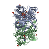
| Title | STRUCTURE OF DIHYDROLIPOAMIDE DEHYDROGENASE | ||||||
 Components Components | SURFACE PROTEIN | ||||||
 Keywords Keywords | OXIDOREDUCTASE / REDOX-ACTIVE CENTER / GLYCOLYSIS / NAD / FLAVOPROTEIN / FAD / P64K | ||||||
| Function / homology |  Function and homology information Function and homology informationdihydrolipoyl dehydrogenase / dihydrolipoyl dehydrogenase (NADH) activity / 2-oxoglutarate metabolic process / flavin adenine dinucleotide binding Similarity search - Function | ||||||
| Biological species |  Neisseria meningitidis (bacteria) Neisseria meningitidis (bacteria) | ||||||
| Method |  X-RAY DIFFRACTION / SYNCHROTRON / MOLECULAR REPLACEMENT, MIR / Resolution: 2.75 Å X-RAY DIFFRACTION / SYNCHROTRON / MOLECULAR REPLACEMENT, MIR / Resolution: 2.75 Å | ||||||
 Authors Authors | Li De La Sierra, I. / Prange, T. / Pernot, L. | ||||||
 Citation Citation | Journal: J.Mol.Biol. / Year: 1997 Title: Molecular structure of the lipoamide dehydrogenase domain of a surface antigen from Neisseria meningitidis. Authors: Li de la Sierra, I. / Pernot, L. / Prange, T. / Saludjian, P. / Schiltz, M. / Fourme, R. / Padron, G. #1: Journal: J.Mol.Biol. / Year: 1994Title: Crystallization and Preliminary X-Ray Investigation of a Recombinant Outer Membrane Protein from Neisseria Meningitidis Authors: Li De La Sierra, I. / Prange, T. / Fourme, R. / Padron, G. / Fuentes, P. / Musacchio, A. / Madrazo, J. #2: Journal: Proteins / Year: 1992Title: The Refined Crystal Structure of Pseudomonas Putida Lipoamide Dehydrogenase Complexed with Nad+ at 2.45 A Resolution Authors: Mattevi, A. / Obmolova, G. / Sokatch, J.R. / Betzel, C. / Hol, W.G. #3: Journal: J.Mol.Biol. / Year: 1991Title: Refined Crystal Structure of Lipoamide Dehydrogenase from Azotobacter Vinelandii at 2.2 A Resolution. A Comparison with the Structure of Glutathione Reductase Authors: Mattevi, A. / Schierbeek, A.J. / Hol, W.G. | ||||||
| History |
|
- Structure visualization
| Structure viewer | Molecule: MolmilJmol/JSmol |
|---|
- Downloads & links
-Download
| PDBx/mmCIF format | 1ojt.cif.gz | 109 KB | Display | PDBx/mmCIF format |
|---|---|---|---|---|
| PDB format | pdb1ojt.ent.gz | 81.4 KB | Display | PDB format |
| PDBx/mmJSON format | 1ojt.json.gz | Tree view | PDBx/mmJSON format | |
| Others |  Other downloads Other downloads |
-Validation report
| Arichive directory | https://data.pdbj.org/pub/pdb/validation_reports/oj/1ojtftp://data.pdbj.org/pub/pdb/validation_reports/oj/1ojt | HTTPS FTP |
|---|
-Related structure data
| Related structure data |  1lvlS S: Starting model for refinement |
|---|---|
| Similar structure data |


-Links
 PDBj
PDBj


- Assembly
| Deposited unit | 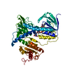
| ||||||||
|---|---|---|---|---|---|---|---|---|---|
| 1 | 
| ||||||||
| Unit cell |
|
-Components
| #1: Protein | Mass: 50737.961 Da / Num. of mol.: 1 / Fragment: DIHYDROLIPOAMIDE DEHYDROGENASE DOMAIN Source method: isolated from a genetically manipulated source Source: (gene. exp.) Neisseria meningitidis (bacteria) / Gene: M-6 OBTAINED FROM A GENOMIC / Gene (production host): M-6 OBTAINED FROM A GENOMIC / Production host: |
|---|---|
| #2: Chemical | ChemComp-FAD /   Mass: 785.550 Da / Num. of mol.: 1 / Source method: obtained synthetically / Formula: C27H33N9O15P2 / Comment: FAD*YM Mass: 785.550 Da / Num. of mol.: 1 / Source method: obtained synthetically / Formula: C27H33N9O15P2 / Comment: FAD*YM |
| #3: Water | ChemComp-HOH /  Mass: 18.015 Da / Num. of mol.: 115 / Source method: isolated from a natural source / Formula: H2O Mass: 18.015 Da / Num. of mol.: 115 / Source method: isolated from a natural source / Formula: H2O |
| Compound details | THE ORIGINAL RECOMBINANT PROTEIN CONTAINS THE TWO DOMAINS E2 AND E3 OF THE DEHYDROGENASE ...THE ORIGINAL RECOMBINAN |
| Has protein modification | Y |
-Experimental details
-Experiment
| Experiment | Method: X-RAY DIFFRACTION / Number of used crystals: 3 |
|---|
- Sample preparation
| Crystal | Density Matthews: 2.73 Å3/Da / Density % sol: 55 % Description: MIR USED 2 DERIVATIVES, ONE MERCURY (TAMM) IN 3 SITES, AND ONE XENON (PRESSURE = 15 BAR) IN 2 SITES. THE TWO MR AND MIR MAPS WERE COMBINED WITH SIGMAA PROGRAM (CCP4) AND SOLVENT ...Description: MIR USED 2 DERIVATIVES, ONE MERCURY (TAMM) IN 3 SITES, AND ONE XENON (PRESSURE = 15 BAR) IN 2 SITES. THE TWO MR AND MIR MAPS WERE COMBINED WITH SIGMAA PROGRAM (CCP4) AND SOLVENT FLATTENED WITH DM (CCP4) AND SOLOMON PROGRAMS. | ||||||||||||||||||||||||||||||||||||||||||
|---|---|---|---|---|---|---|---|---|---|---|---|---|---|---|---|---|---|---|---|---|---|---|---|---|---|---|---|---|---|---|---|---|---|---|---|---|---|---|---|---|---|---|---|
| Crystal grow | Method: vapor diffusion, hanging drop / pH: 7 Details: BRIGHT YELLOW CRYSTALS WERE GROWN IN 24-WELL LINBRO PLATES USING HANGING DROP DIFFUSION METHOD AT ROOM TEMPERATURE. RESERVOIR: 1 ML CONTAINING 0.1M POTASSIUM PHOSPHATE AND 2M AMMONIUM ...Details: BRIGHT YELLOW CRYSTALS WERE GROWN IN 24-WELL LINBRO PLATES USING HANGING DROP DIFFUSION METHOD AT ROOM TEMPERATURE. RESERVOIR: 1 ML CONTAINING 0.1M POTASSIUM PHOSPHATE AND 2M AMMONIUM SULFATE (PH 7.0). PRISM CRYSTALS TYPICALLY IN TWO WEEKS AT ROOM TEMPERATURE., vapor diffusion - hanging drop THE E3 DOMAIN (117-601) CRYSTALLIZES IN AMMONIUM SULFATE. IT INCLUDES A FAD COFACTOR BUT NOT THE NADH. THE E3 DOMAIN (117-601) CRYSTALLIZES IN AMMONIUM SULFATE. IT INCLUDE A FAD COFACTOR BUT NOT THE NADH. Temp details: room temp | ||||||||||||||||||||||||||||||||||||||||||
| Crystal grow | *PLUS pH: 7.2 / Method: vapor diffusion, hanging dropDetails: Li De La Sierra, I., (1994) J.Mol.Biol., 235, 1154. | ||||||||||||||||||||||||||||||||||||||||||
| Components of the solutions | *PLUS
|
-Data collection
| Diffraction | Mean temperature: 277 K |
|---|---|
| Diffraction source | Source: SYNCHROTRON / Site: LURE  / Type: LURE / Wavelength: 0.91 / Type: LURE / Wavelength: 0.91 |
| Detector | Type: MAR scanner 300 mm plate / Detector: IMAGE PLATE / Details: MULTILAYER MIRROR |
| Radiation | Monochromator: SI(111) / Monochromatic (M) / Laue (L): M / Scattering type: x-ray |
| Radiation wavelength | Wavelength: 0.91 Å / Relative weight: 1 |
| Reflection | Resolution: 2.7→40 Å / Num. obs: 11724 / % possible obs: 94 % / Observed criterion σ(I): 3 / Redundancy: 7 % / Rmerge(I) obs: 0.066 / Net I/σ(I): 18 |
| Reflection shell | Resolution: 2.7→2.9 Å / Redundancy: 4 % / Rmerge(I) obs: 0.19 / Mean I/σ(I) obs: 4 / % possible all: 70 |
- Processing
| Software |
| |||||||||||||||||||||||||||||||||||||||||||||||||||||||||||||||
|---|---|---|---|---|---|---|---|---|---|---|---|---|---|---|---|---|---|---|---|---|---|---|---|---|---|---|---|---|---|---|---|---|---|---|---|---|---|---|---|---|---|---|---|---|---|---|---|---|---|---|---|---|---|---|---|---|---|---|---|---|---|---|---|---|
| Refinement | Method to determine structure: MOLECULAR REPLACEMENT, MIR Starting model: DLDH OF PSEUDOMONAS PUTIDA, PDB ENTRY 1LVL. Resolution: 2.75→18 Å / Details: X-PLOR ALSO WAS USED. /
| |||||||||||||||||||||||||||||||||||||||||||||||||||||||||||||||
| Refinement step | Cycle: LAST / Resolution: 2.75→18 Å
| |||||||||||||||||||||||||||||||||||||||||||||||||||||||||||||||
| Refine LS restraints |
| |||||||||||||||||||||||||||||||||||||||||||||||||||||||||||||||
| Software | *PLUS Name: PROLSQ / Classification: refinement | |||||||||||||||||||||||||||||||||||||||||||||||||||||||||||||||
| Refinement | *PLUS Lowest resolution: 5 Å / Rfactor all: 0.201 | |||||||||||||||||||||||||||||||||||||||||||||||||||||||||||||||
| Solvent computation | *PLUS | |||||||||||||||||||||||||||||||||||||||||||||||||||||||||||||||
| Displacement parameters | *PLUS |
