 Movie
Movie Controller

[English] 日本語
 Yorodumi
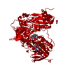
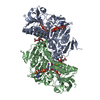
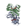
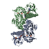
- PDB-4new: Crystal structure of Trypanothione Reductase from Trypanosoma cru... -
+ Open data

- Basic information
| Entry | Database: PDB / ID: 4new | ||||||
|---|---|---|---|---|---|---|---|
| Title | Crystal structure of Trypanothione Reductase from Trypanosoma cruzi in complex with inhibitor EP127 (5-{5-[1-(PYRROLIDIN-1-YL)CYCLOHEXYL]-1,3-THIAZOL-2-YL}-1H-INDOLE) | ||||||
 Components Components | Trypanothione reductase, putative | ||||||
 Keywords Keywords | Oxidoreductase/Oxidoreductase inhibitor / Reductase / Oxidoreductase-Oxidoreductase inhibitor complex | ||||||
| Function / homology |  Function and homology information Function and homology informationFAD/NAD-linked reductase, C-terminal dimerisation domain / Enolase-like; domain 1 / FAD/NAD(P)-binding domain / FAD/NAD(P)-binding domain / 3-Layer(bba) Sandwich / 2-Layer Sandwich / Alpha Beta Similarity search - Domain/homology | ||||||
| Biological species |  | ||||||
| Method |  X-RAY DIFFRACTION / MOLECULAR REPLACEMENT / Resolution: 2.8 Å X-RAY DIFFRACTION / MOLECULAR REPLACEMENT / Resolution: 2.8 Å | ||||||
 Authors Authors | Persch, E. / Bryson, S. / Pai, E.F. / Krauth-Siegel, R.L. / Diederich, F. | ||||||
 Citation Citation | Journal: Chemmedchem / Year: 2014 Title: Binding to large enzyme pockets: small-molecule inhibitors of trypanothione reductase. Authors: Persch, E. / Bryson, S. / Todoroff, N.K. / Eberle, C. / Thelemann, J. / Dirdjaja, N. / Kaiser, M. / Weber, M. / Derbani, H. / Brun, R. / Schneider, G. / Pai, E.F. / Krauth-Siegel, R.L. / Diederich, F. | ||||||
| History |
|
- Structure visualization
| Structure viewer | Molecule: MolmilJmol/JSmol |
|---|
- Downloads & links
-Download
| PDBx/mmCIF format | 4new.cif.gz | 108.6 KB | Display | PDBx/mmCIF format |
|---|---|---|---|---|
| PDB format | pdb4new.ent.gz | 82.4 KB | Display | PDB format |
| PDBx/mmJSON format | 4new.json.gz | Tree view | PDBx/mmJSON format | |
| Others |  Other downloads Other downloads |
-Validation report
| Arichive directory | https://data.pdbj.org/pub/pdb/validation_reports/ne/4newftp://data.pdbj.org/pub/pdb/validation_reports/ne/4new | HTTPS FTP |
|---|
-Related structure data
| Related structure data |  4nevC  1bzlS C: citing same article ( S: Starting model for refinement |
|---|---|
| Similar structure data |


-Links
 PDBj
PDBj- Assembly
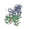
| Deposited unit | 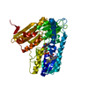
| ||||||||
|---|---|---|---|---|---|---|---|---|---|
| 1 | 
| ||||||||
| Unit cell |
|
-Components
| #1: Protein | Mass: 53935.703 Da / Num. of mol.: 1 Source method: isolated from a genetically manipulated source Source: (gene. exp.)  |
|---|---|
| #2: Chemical | ChemComp-FAD /   Mass: 785.550 Da / Num. of mol.: 1 / Source method: obtained synthetically / Formula: C27H33N9O15P2 / Comment: FAD*YM Mass: 785.550 Da / Num. of mol.: 1 / Source method: obtained synthetically / Formula: C27H33N9O15P2 / Comment: FAD*YM |
| #3: Chemical | ChemComp-2JR /   Mass: 351.508 Da / Num. of mol.: 1 / Source method: obtained synthetically / Formula: C21H25N3S Mass: 351.508 Da / Num. of mol.: 1 / Source method: obtained synthetically / Formula: C21H25N3S |
| Has protein modification | Y |
| Sequence details | AUTHORS HAVE INDICATED THAT THE RESIDUES ASN95 COULD NOT BE CLEARLY IDENTIFIED FROM THE ...AUTHORS HAVE INDICATED THAT THE RESIDUES ASN95 COULD NOT BE CLEARLY IDENTIFIED |
-Experimental details
-Experiment
| Experiment | Method: X-RAY DIFFRACTION / Number of used crystals: 1 |
|---|
- Sample preparation
| Crystal | Density Matthews: 2.64 Å3/Da / Density % sol: 53.44 % |
|---|---|
| Crystal grow | Temperature: 298 K / pH: 8 Details: 0.1M TRIS, pH 8.0, 0.2M CaCl2, 20-22% PEG2000MME, VAPOR DIFFUSION, HANGING DROP, temperature 298K |
-Data collection
| Diffraction | Mean temperature: 120 K |
|---|---|
| Diffraction source | Source: ROTATING ANODE / Type: RIGAKU MICROMAX-007 HF / Wavelength: 1.5418 |
| Detector | Type: MAR scanner 345 mm plate / Detector: IMAGE PLATE / Date: Apr 9, 2013 |
| Radiation | Monochromator: NI FILTER / Protocol: SINGLE WAVELENGTH / Monochromatic (M) / Laue (L): M / Scattering type: x-ray |
| Radiation wavelength | Wavelength: 1.5418 Å / Relative weight: 1 |
| Reflection | Resolution: 2.8→17 Å / Num. obs: 14005 / % possible obs: 93.9 % / Observed criterion σ(I): 0 / Redundancy: 1.72 % / Rmerge(I) obs: 0.028 |
| Reflection shell | Resolution: 2.8→2.9 Å / Rmerge(I) obs: 0.11 / Mean I/σ(I) obs: 7.3 / % possible all: 75.4 |
- Processing
| Software |
| ||||||||||||||||||
|---|---|---|---|---|---|---|---|---|---|---|---|---|---|---|---|---|---|---|---|
| Refinement | Method to determine structure: MOLECULAR REPLACEMENT Starting model: 1BZL Resolution: 2.8→17 Å / σ(F): 0 / Stereochemistry target values: MAXIMUM LIKELIHOOD
| ||||||||||||||||||
| Refinement step | Cycle: LAST / Resolution: 2.8→17 Å
| ||||||||||||||||||
| Refine LS restraints |
|
