 Movie
Movie Controller

[English] 日本語
 Yorodumi
- PDB-6bu7: Crystal structure of Trypanothione Reductase from Trypanosoma bru... -
+ Open data

- Basic information
| Entry | Database: PDB / ID: 6bu7 | |||||||||||||||
|---|---|---|---|---|---|---|---|---|---|---|---|---|---|---|---|---|
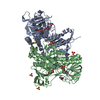
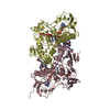
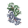
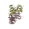
| Title | Crystal structure of Trypanothione Reductase from Trypanosoma brucei in complex with inhibitor RD130 1-[2-(Piperidin-4-yl)ethyl]-5-{5-[1-(pyrrolidin-1-yl)cyclohexyl]-1,3-thiazol-2-yl}-1H-indole | |||||||||||||||
 Components Components | Trypanothione reductase | |||||||||||||||
 Keywords Keywords | Oxidoreductase/Inhibitor / Trypanosoma / Inhibitor / Complex / Sleeping Sickness / Oxidoreductase-Inhibitor complex | |||||||||||||||
| Function / homology |  Function and homology information Function and homology informationtrypanothione-disulfide reductase / trypanothione-disulfide reductase (NADPH) activity / kinetoplast / glycosome / nuclear lumen / thioredoxin-disulfide reductase (NADPH) activity / ciliary plasm / cell redox homeostasis / flavin adenine dinucleotide binding / nucleoplasm ...trypanothione-disulfide reductase / trypanothione-disulfide reductase (NADPH) activity / kinetoplast / glycosome / nuclear lumen / thioredoxin-disulfide reductase (NADPH) activity / ciliary plasm / cell redox homeostasis / flavin adenine dinucleotide binding / nucleoplasm / metal ion binding / cytoplasm Similarity search - Function | |||||||||||||||
| Biological species |  | |||||||||||||||
| Method |  X-RAY DIFFRACTION / SYNCHROTRON / MOLECULAR REPLACEMENT / Resolution: 2.73 Å X-RAY DIFFRACTION / SYNCHROTRON / MOLECULAR REPLACEMENT / Resolution: 2.73 Å | |||||||||||||||
 Authors Authors | Bryson, S. / De Gasparo, R. / Krauth-Siegel, R.L. / Diederich, F. / Pai, E.F. | |||||||||||||||
| Funding support |  Canada, Canada,  Switzerland, Switzerland,  Germany, 4items Germany, 4items
| |||||||||||||||
 Citation Citation | Journal: ChemMedChem / Year: 2018 Title: Biological Evaluation and X-ray Co-crystal Structures of Cyclohexylpyrrolidine Ligands for Trypanothione Reductase, an Enzyme from the Redox Metabolism of Trypanosoma. Authors: De Gasparo, R. / Brodbeck-Persch, E. / Bryson, S. / Hentzen, N.B. / Kaiser, M. / Pai, E.F. / Krauth-Siegel, R.L. / Diederich, F. #1: Journal: ChemMedChem / Year: 2014Title: Binding to large enzyme pockets: small-molecule inhibitors of trypanothione reductase. Authors: Persch, E. / Bryson, S. / Todoroff, N.K. / Eberle, C. / Thelemann, J. / Dirdjaja, N. / Kaiser, M. / Weber, M. / Derbani, H. / Brun, R. / Schneider, G. / Pai, E.F. / Krauth-Siegel, R.L. / Diederich, F. | |||||||||||||||
| History |
|
- Structure visualization
| Structure viewer | Molecule: MolmilJmol/JSmol |
|---|
- Downloads & links
-Download
| PDBx/mmCIF format | 6bu7.cif.gz | 406.8 KB | Display | PDBx/mmCIF format |
|---|---|---|---|---|
| PDB format | pdb6bu7.ent.gz | 331.2 KB | Display | PDB format |
| PDBx/mmJSON format | 6bu7.json.gz | Tree view | PDBx/mmJSON format | |
| Others |  Other downloads Other downloads |
-Validation report
| Arichive directory | https://data.pdbj.org/pub/pdb/validation_reports/bu/6bu7ftp://data.pdbj.org/pub/pdb/validation_reports/bu/6bu7 | HTTPS FTP |
|---|
-Related structure data
| Related structure data |  6btlC  2woiS C: citing same article ( S: Starting model for refinement |
|---|---|
| Similar structure data |


-Links
 PDBj
PDBj


- Assembly
| Deposited unit | 
| ||||||||
|---|---|---|---|---|---|---|---|---|---|
| 1 |
| ||||||||
| Unit cell |
|
-Components
-Protein , 1 types, 2 molecules AB
| #1: Protein | Mass: 53497.969 Da / Num. of mol.: 2 Source method: isolated from a genetically manipulated source Source: (gene. exp.) Strain: 927/4 GUTat10.1 / Gene: Tb10.406.0520 / Plasmid: pET3aTbTryR Production host:  References: UniProt: Q389T8, trypanothione-disulfide reductase |
|---|
-Non-polymers , 6 types, 34 molecules 


| #2: Chemical |  Mass: 785.550 Da / Num. of mol.: 2 / Source method: obtained synthetically / Formula: C27H33N9O15P2 / Comment: FAD*YM Mass: 785.550 Da / Num. of mol.: 2 / Source method: obtained synthetically / Formula: C27H33N9O15P2 / Comment: FAD*YM#3: Chemical | ChemComp-SO4 /  Mass: 96.063 Da / Num. of mol.: 5 / Source method: obtained synthetically / Formula: SO4 Mass: 96.063 Da / Num. of mol.: 5 / Source method: obtained synthetically / Formula: SO4#4: Chemical |  Mass: 462.693 Da / Num. of mol.: 2 / Source method: obtained synthetically / Formula: C28H38N4S / Feature type: SUBJECT OF INVESTIGATION Mass: 462.693 Da / Num. of mol.: 2 / Source method: obtained synthetically / Formula: C28H38N4S / Feature type: SUBJECT OF INVESTIGATION#5: Chemical |  Mass: 238.305 Da / Num. of mol.: 2 / Source method: obtained synthetically / Formula: C8H18N2O4S / Comment: pH buffer*YM Mass: 238.305 Da / Num. of mol.: 2 / Source method: obtained synthetically / Formula: C8H18N2O4S / Comment: pH buffer*YM#6: Chemical | ChemComp-GOL /  Mass: 92.094 Da / Num. of mol.: 4 / Source method: obtained synthetically / Formula: C3H8O3 Mass: 92.094 Da / Num. of mol.: 4 / Source method: obtained synthetically / Formula: C3H8O3#7: Water | ChemComp-HOH / | Mass: 18.015 Da / Num. of mol.: 19 / Source method: isolated from a natural source / Formula: H2O |
|---|
-Details
| Has protein modification | Y |
|---|
-Experimental details
-Experiment
| Experiment | Method: X-RAY DIFFRACTION / Number of used crystals: 1 |
|---|
- Sample preparation
| Crystal | Density Matthews: 3.61 Å3/Da / Density % sol: 65.91 % |
|---|---|
| Crystal grow | Temperature: 294 K / Method: vapor diffusion, hanging drop / pH: 7.5 Details: Adding 5 microL of 10 mM inhibitor in DMSO to 95 microL of protein solution (10mg/ml; 20 mM TRIS, pH8.0), then mixing 2 microL of protein solution with 2 microL of well solution (0.1 M ...Details: Adding 5 microL of 10 mM inhibitor in DMSO to 95 microL of protein solution (10mg/ml; 20 mM TRIS, pH8.0), then mixing 2 microL of protein solution with 2 microL of well solution (0.1 M HEPES, pH 7.5, 2.0 M (NH4)2SO4). |
-Data collection
| Diffraction | Mean temperature: 93 K |
|---|---|
| Diffraction source | Source: SYNCHROTRON / Site: APS  / Beamline: 23-ID-D / Wavelength: 1.0332 Å / Beamline: 23-ID-D / Wavelength: 1.0332 Å |
| Detector | Type: DECTRIS PILATUS3 6M / Detector: PIXEL / Date: Dec 3, 2016 / Details: collimator |
| Radiation | Monochromator: double crystal monochromator / Protocol: SINGLE WAVELENGTH / Monochromatic (M) / Laue (L): M / Scattering type: x-ray |
| Radiation wavelength | Wavelength: 1.0332 Å / Relative weight: 1 |
| Reflection | Resolution: 2.73→46.5 Å / Num. obs: 42552 / % possible obs: 99.9 % / Redundancy: 8.8 % / Biso Wilson estimate: 74.1 Å2 / CC1/2: 0.999 / Rmerge(I) obs: 0.104 / Rpim(I) all: 0.037 / Rrim(I) all: 0.111 / Net I/σ(I): 12.6 |
| Reflection shell | Resolution: 2.73→2.82 Å / Redundancy: 7.3 % / Rmerge(I) obs: 1.469 / Mean I/σ(I) obs: 1.04 / Num. unique obs: 4132 / CC1/2: 0.587 / Rpim(I) all: 0.569 / Rrim(I) all: 1.583 / % possible all: 99.9 |
- Processing
| Software |
| ||||||||||||||||||||||||||||||||||||||||||||||||||||||||||||||||||||||||||||||||||||||||||||||||||||||||||||||||
|---|---|---|---|---|---|---|---|---|---|---|---|---|---|---|---|---|---|---|---|---|---|---|---|---|---|---|---|---|---|---|---|---|---|---|---|---|---|---|---|---|---|---|---|---|---|---|---|---|---|---|---|---|---|---|---|---|---|---|---|---|---|---|---|---|---|---|---|---|---|---|---|---|---|---|---|---|---|---|---|---|---|---|---|---|---|---|---|---|---|---|---|---|---|---|---|---|---|---|---|---|---|---|---|---|---|---|---|---|---|---|---|---|---|
| Refinement | Method to determine structure: MOLECULAR REPLACEMENT Starting model: 2WOI Resolution: 2.73→46.5 Å / SU ML: 0.43 / Cross valid method: FREE R-VALUE / σ(F): 1.34 / Phase error: 28.82
| ||||||||||||||||||||||||||||||||||||||||||||||||||||||||||||||||||||||||||||||||||||||||||||||||||||||||||||||||
| Solvent computation | Shrinkage radii: 0.9 Å / VDW probe radii: 1.11 Å | ||||||||||||||||||||||||||||||||||||||||||||||||||||||||||||||||||||||||||||||||||||||||||||||||||||||||||||||||
| Displacement parameters | Biso mean: 84.7 Å2 | ||||||||||||||||||||||||||||||||||||||||||||||||||||||||||||||||||||||||||||||||||||||||||||||||||||||||||||||||
| Refinement step | Cycle: LAST / Resolution: 2.73→46.5 Å
| ||||||||||||||||||||||||||||||||||||||||||||||||||||||||||||||||||||||||||||||||||||||||||||||||||||||||||||||||
| Refine LS restraints |
| ||||||||||||||||||||||||||||||||||||||||||||||||||||||||||||||||||||||||||||||||||||||||||||||||||||||||||||||||
| LS refinement shell |
| ||||||||||||||||||||||||||||||||||||||||||||||||||||||||||||||||||||||||||||||||||||||||||||||||||||||||||||||||
| Refinement TLS params. | Method: refined / Origin x: 31.8884 Å / Origin y: -13.0406 Å / Origin z: 6.9739 Å
| ||||||||||||||||||||||||||||||||||||||||||||||||||||||||||||||||||||||||||||||||||||||||||||||||||||||||||||||||
| Refinement TLS group | Selection details: all |
