[English] 日本語
Yorodumi
- PDB-2wba: Properties of Trypanothione Reductase From T. brucei -

+
Open data


ID or keywords:

Loading...

-
Basic information

Entry
Database: PDB / ID: 2wba
TitleProperties of Trypanothione Reductase From T. brucei
ComponentsTRYPANOTHIONE REDUCTASE
KeywordsOXIDOREDUCTASE / REDOX-ACTIVE CENTER / FLAVOPROTEIN / TRYPNOTHIONE METABOLISM OXIDOREDUCTASE
Function / homology
Function and homology information


trypanothione-disulfide reductase / trypanothione-disulfide reductase (NADPH) activity / glutathione-disulfide reductase (NADPH) activity / glutathione metabolic process / cell redox homeostasis / flavin adenine dinucleotide binding / cellular response to oxidative stress / mitochondrion / cytosol
Similarity search - Function
Trypanothione reductase / : / Pyridine nucleotide-disulphide oxidoreductase, class I / FAD/NAD-linked reductase, C-terminal dimerisation domain / Pyridine nucleotide-disulphide oxidoreductase, class I, active site / Pyridine nucleotide-disulphide oxidoreductases class-I active site. / Pyridine nucleotide-disulphide oxidoreductase, dimerisation domain / Pyridine nucleotide-disulphide oxidoreductase, dimerisation domain / FAD/NAD-linked reductase, dimerisation domain superfamily / FAD/NAD(P)-binding domain ...Trypanothione reductase / : / Pyridine nucleotide-disulphide oxidoreductase, class I / FAD/NAD-linked reductase, C-terminal dimerisation domain / Pyridine nucleotide-disulphide oxidoreductase, class I, active site / Pyridine nucleotide-disulphide oxidoreductases class-I active site. / Pyridine nucleotide-disulphide oxidoreductase, dimerisation domain / Pyridine nucleotide-disulphide oxidoreductase, dimerisation domain / FAD/NAD-linked reductase, dimerisation domain superfamily / FAD/NAD(P)-binding domain / Pyridine nucleotide-disulphide oxidoreductase / Enolase-like; domain 1 / FAD/NAD(P)-binding domain / FAD/NAD(P)-binding domain / 3-Layer(bba) Sandwich / FAD/NAD(P)-binding domain superfamily / 2-Layer Sandwich / Alpha Beta
Similarity search - Domain/homology
FLAVIN-ADENINE DINUCLEOTIDE / Chem-NDP / Trypanothione reductase
Similarity search - Component
Biological speciesTRYPANOSOMA BRUCEI BRUCEI (eukaryote)
MethodX-RAY DIFFRACTION / MOLECULAR REPLACEMENT / Resolution: 2.3 Å
AuthorsJones, D. / Ariza, A. / Chow, W.H.A. / Oza, S.L. / Fairlamb, A.H.
CitationJournal: Mol.Biochem.Parasitol. / Year: 2010
Title: Comparative Structural, Kinetic and Inhibitor Studies of Trypanosoma Brucei Trypanothione Reductase with T. Cruzi.
Authors: Jones, D. / Ariza, A. / Chow, W.H.A. / Oza, S.L. / Fairlamb, A.H.
History
DepositionFeb 24, 2009Deposition site: PDBE / Processing site: PDBE
Revision 1.0Nov 24, 2009Provider: repository / Type: Initial release
SupersessionMay 19, 2010ID: 2VE2
Revision 1.1May 8, 2011Group: Version format compliance
Revision 1.2Jul 13, 2011Group: Version format compliance
Revision 1.3Dec 13, 2023Group: Data collection / Database references ...Data collection / Database references / Derived calculations / Other / Refinement description
Category: chem_comp_atom / chem_comp_bond ...chem_comp_atom / chem_comp_bond / database_2 / pdbx_database_status / pdbx_initial_refinement_model / struct_site
Item: _database_2.pdbx_DOI / _database_2.pdbx_database_accession ..._database_2.pdbx_DOI / _database_2.pdbx_database_accession / _pdbx_database_status.status_code_sf / _struct_site.pdbx_auth_asym_id / _struct_site.pdbx_auth_comp_id / _struct_site.pdbx_auth_seq_id
Remark 700 
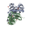
SHEET THE SHEET STRUCTURE OF THIS MOLECULE IS BIFURCATED. IN ORDER TO REPRESENT THIS FEATURE IN ... SHEET THE SHEET STRUCTURE OF THIS MOLECULE IS BIFURCATED. IN ORDER TO REPRESENT THIS FEATURE IN THE SHEET RECORDS BELOW, TWO SHEETS ARE DEFINED.

-
Structure visualization

Structure viewerMolecule:
MolmilJmol/JSmol

Downloads & links

-
Assembly

Deposited unit
A: TRYPANOTHIONE REDUCTASE
B: TRYPANOTHIONE REDUCTASE
hetero molecules


Theoretical massNumber of molelcules
Total (without water)110,61617
Polymers106,4312
Non-polymers4,18515
Water14,160786
1


  • Idetical with deposited unit
  • defined by author&software
TypeNameSymmetry operationNumber
identity operation1_555x,y,z1
Buried area13350 Å2
ΔGint-90.5 kcal/mol
Surface area37170 Å2
MethodPISA
Unit cell
Length a, b, c (Å)63.630, 132.713, 161.287
Angle α, β, γ (deg.)90.00, 90.00, 90.00
Int Tables number19
Space group name H-MP212121

-
Components

-
Protein , 1 types, 2 molecules AB

#1: Protein TRYPANOTHIONE REDUCTASE


Mass: 53215.688 Da / Num. of mol.: 2
Source method: isolated from a genetically manipulated source
Source: (gene. exp.) TRYPANOSOMA BRUCEI BRUCEI (eukaryote) / Strain: S427 / Plasmid: PET3A / Production host: ESCHERICHIA COLI (E. coli) / Strain (production host): BL21 STAR (DE3) PLYSS
References: UniProt: P39051*PLUS, trypanothione-disulfide reductase

-
Non-polymers , 6 types, 801 molecules

#2: Chemical ChemComp-FAD / FLAVIN-ADENINE DINUCLEOTIDE


Mass: 785.550 Da / Num. of mol.: 2 / Source method: obtained synthetically / Formula: C27H33N9O15P2 / Comment: FAD*YM
#3: Chemical
ChemComp-GOL / GLYCEROL / GLYCERIN / PROPANE-1,2,3-TRIOL


Mass: 92.094 Da / Num. of mol.: 8 / Source method: obtained synthetically / Formula: C3H8O3
#4: Chemical ChemComp-NDP / NADPH DIHYDRO-NICOTINAMIDE-ADENINE-DINUCLEOTIDE PHOSPHATE


Mass: 745.421 Da / Num. of mol.: 2 / Source method: obtained synthetically / Formula: C21H30N7O17P3
#5: Chemical ChemComp-PG4 / TETRAETHYLENE GLYCOL


Mass: 194.226 Da / Num. of mol.: 1 / Source method: obtained synthetically / Formula: C8H18O5 / Comment: precipitant*YM
#6: Chemical ChemComp-SO4 / SULFATE ION


Mass: 96.063 Da / Num. of mol.: 2 / Source method: obtained synthetically / Formula: SO4
#7: Water ChemComp-HOH / water


Mass: 18.015 Da / Num. of mol.: 786 / Source method: isolated from a natural source / Formula: H2O

-
Details

Sequence detailsTHE ORF WAS IDENTIFIED FROM A PUTATIVE TRYPANOTHIONE REDUCTASE FROM T. BRUCEI STRAIN S427 IN THE ...THE ORF WAS IDENTIFIED FROM A PUTATIVE TRYPANOTHIONE REDUCTASE FROM T. BRUCEI STRAIN S427 IN THE GENEDB. THE SAME ORF OUT GB TB10.406.0520 ENTRY (FROM STRAIN S927) WAS CLONED OF A DIFFERENT T. BRUCEI BRUCEI STRAIN (STRAIN S427). THE ORF OF THE PROTEIN IS NEARLY IDENTICAL TO THE TB10.406.0520 ORF (THERE ARE ONLY 2 NUCLEOTIDES THAT ARE DIFFERENT OUT OF 1479 BASE PAIRS AND NEITHER OF THESE CHANGE THE PROTEIN SEQUENCE, WHICH IS 100% IDENTICAL BETWEEN BOTH STRAINS: S927 AND S427

-
Experimental details

-
Experiment

ExperimentMethod: X-RAY DIFFRACTION / Number of used crystals: 1

-
Sample preparation

CrystalDensity Matthews: 3.2 Å3/Da / Density % sol: 62 % / Description: NONE
Crystal growpH: 8
Details: 0.1 M BIS-TRIS PH 8.0, 2.0 AMMONIUM SULFATE, 5 % PEG 400

-
Data collection

DiffractionMean temperature: 100 K
Diffraction sourceSource: ROTATING ANODE / Type: RIGAKU MICROMAX-007 HF / Wavelength: 1.5428
DetectorType: RIGAKU IMAGE PLATE / Detector: IMAGE PLATE / Date: Aug 28, 2006 / Details: MIRRORS (OSMIC BLUE)
RadiationProtocol: SINGLE WAVELENGTH / Monochromatic (M) / Laue (L): M / Scattering type: x-ray
Radiation wavelengthWavelength: 1.5428 Å / Relative weight: 1
ReflectionResolution: 2.3→41.07 Å / Num. obs: 55415 / % possible obs: 94.9 % / Observed criterion σ(I): 0 / Redundancy: 5 % / Rmerge(I) obs: 0.05 / Net I/σ(I): 22.9
Reflection shellResolution: 2.3→2.42 Å / Redundancy: 3.4 % / Rmerge(I) obs: 0.1 / Mean I/σ(I) obs: 9.2 / % possible all: 85.7

-
Processing

Software
NameVersionClassification
REFMAC5.5.0082refinement
MOSFLMdata reduction
SCALAdata scaling
MOLREPphasing
RefinementMethod to determine structure: MOLECULAR REPLACEMENT
Starting model: PDB ENTRY 1BZL
Resolution: 2.3→102.6 Å / Cor.coef. Fo:Fc: 0.939 / Cor.coef. Fo:Fc free: 0.907 / SU B: 5.072 / SU ML: 0.127 / Cross valid method: THROUGHOUT / ESU R: 0.273 / ESU R Free: 0.213 / Stereochemistry target values: MAXIMUM LIKELIHOOD
Details: HYDROGENS HAVE BEEN ADDED IN THE RIDING POSITIONS. U VALUES REFINED INDIVIDUALLY
RfactorNum. reflection% reflectionSelection details
Rfree0.22715 2952 5.1 %RANDOM
Rwork0.1808 ---
obs0.18315 55398 94.81 %-
Solvent computationIon probe radii: 0.8 Å / Shrinkage radii: 0.8 Å / VDW probe radii: 1.4 Å / Solvent model: MASK
Displacement parametersBiso mean: 19.666 Å2
Baniso -1Baniso -2Baniso -3
1-1.11 Å20 Å20 Å2
2---1.04 Å20 Å2
3----0.07 Å2
Refinement stepCycle: LAST / Resolution: 2.3→102.6 Å
ProteinNucleic acidLigandSolventTotal
Num. atoms7424 0 235 786 8445
Refine LS restraints
Refine-IDTypeDev idealDev ideal targetNumber
X-RAY DIFFRACTIONr_bond_refined_d0.0040.0227935
X-RAY DIFFRACTIONr_bond_other_d
X-RAY DIFFRACTIONr_angle_refined_deg0.9561.99610806
X-RAY DIFFRACTIONr_angle_other_deg
X-RAY DIFFRACTIONr_dihedral_angle_1_deg5.67351006
X-RAY DIFFRACTIONr_dihedral_angle_2_deg34.0124.591318
X-RAY DIFFRACTIONr_dihedral_angle_3_deg13.678151299
X-RAY DIFFRACTIONr_dihedral_angle_4_deg13.0891537
X-RAY DIFFRACTIONr_chiral_restr0.0740.21207
X-RAY DIFFRACTIONr_gen_planes_refined0.0050.0215881
X-RAY DIFFRACTIONr_gen_planes_other
X-RAY DIFFRACTIONr_nbd_refined0.1860.23469
X-RAY DIFFRACTIONr_nbd_other
X-RAY DIFFRACTIONr_nbtor_refined0.3050.25410
X-RAY DIFFRACTIONr_nbtor_other
X-RAY DIFFRACTIONr_xyhbond_nbd_refined0.1180.2720
X-RAY DIFFRACTIONr_xyhbond_nbd_other
X-RAY DIFFRACTIONr_metal_ion_refined
X-RAY DIFFRACTIONr_metal_ion_other
X-RAY DIFFRACTIONr_symmetry_vdw_refined0.1580.246
X-RAY DIFFRACTIONr_symmetry_vdw_other
X-RAY DIFFRACTIONr_symmetry_hbond_refined0.1220.217
X-RAY DIFFRACTIONr_symmetry_hbond_other
X-RAY DIFFRACTIONr_symmetry_metal_ion_refined
X-RAY DIFFRACTIONr_symmetry_metal_ion_other
X-RAY DIFFRACTIONr_mcbond_it0.4451.54887
X-RAY DIFFRACTIONr_mcbond_other
X-RAY DIFFRACTIONr_mcangle_it0.86427906
X-RAY DIFFRACTIONr_mcangle_other
X-RAY DIFFRACTIONr_scbond_it1.32133048
X-RAY DIFFRACTIONr_scbond_other
X-RAY DIFFRACTIONr_scangle_it2.2174.52885
X-RAY DIFFRACTIONr_scangle_other
X-RAY DIFFRACTIONr_long_range_B_refined
X-RAY DIFFRACTIONr_long_range_B_other
X-RAY DIFFRACTIONr_rigid_bond_restr
X-RAY DIFFRACTIONr_sphericity_free
X-RAY DIFFRACTIONr_sphericity_bonded
LS refinement shellResolution: 2.3→2.36 Å / Total num. of bins used: 20
RfactorNum. reflection% reflection
Rfree0.29 182 -
Rwork0.217 3563 -
obs--83.58 %

+
About Yorodumi

-
News

-
Feb 9, 2022. New format data for meta-information of EMDB entries

New format data for meta-information of EMDB entries

  • Version 3 of the EMDB header file is now the official format.
  • The previous official version 1.9 will be removed from the archive.

Related info.:EMDB header

External links:wwPDB to switch to version 3 of the EMDB data model

-
Aug 12, 2020. Covid-19 info

Covid-19 info

URL: https://pdbjlvh1.pdbj.org/emnavi/covid19.php

New page: Covid-19 featured information page in EM Navigator.

Related info.:Covid-19 info / Mar 5, 2020. Novel coronavirus structure data

+
Mar 5, 2020. Novel coronavirus structure data

Novel coronavirus structure data

Related info.:Yorodumi Speices / Aug 12, 2020. Covid-19 info

External links:COVID-19 featured content - PDBj / Molecule of the Month (242):Coronavirus Proteases

+
Jan 31, 2019. EMDB accession codes are about to change! (news from PDBe EMDB page)

EMDB accession codes are about to change! (news from PDBe EMDB page)

  • The allocation of 4 digits for EMDB accession codes will soon come to an end. Whilst these codes will remain in use, new EMDB accession codes will include an additional digit and will expand incrementally as the available range of codes is exhausted. The current 4-digit format prefixed with “EMD-” (i.e. EMD-XXXX) will advance to a 5-digit format (i.e. EMD-XXXXX), and so on. It is currently estimated that the 4-digit codes will be depleted around Spring 2019, at which point the 5-digit format will come into force.
  • The EM Navigator/Yorodumi systems omit the EMD- prefix.

Related info.:Q: What is EMD? / ID/Accession-code notation in Yorodumi/EM Navigator

External links:EMDB Accession Codes are Changing Soon! / Contact to PDBj

+
Jul 12, 2017. Major update of PDB

Major update of PDB

  • wwPDB released updated PDB data conforming to the new PDBx/mmCIF dictionary.
  • This is a major update changing the version number from 4 to 5, and with Remediation, in which all the entries are updated.
  • In this update, many items about electron microscopy experimental information are reorganized (e.g. em_software).
  • Now, EM Navigator and Yorodumi are based on the updated data.

External links:wwPDB Remediation / Enriched Model Files Conforming to OneDep Data Standards Now Available in the PDB FTP Archive

-
Yorodumi

Thousand views of thousand structures

  • Yorodumi is a browser for structure data from EMDB, PDB, SASBDB, etc.
  • This page is also the successor to EM Navigator detail page, and also detail information page/front-end page for Omokage search.
  • The word "yorodu" (or yorozu) is an old Japanese word meaning "ten thousand". "mi" (miru) is to see.

Related info.:EMDB / PDB / SASBDB / Comparison of 3 databanks / Yorodumi Search / Aug 31, 2016. New EM Navigator & Yorodumi / Yorodumi Papers / Jmol/JSmol / Function and homology information / Changes in new EM Navigator and Yorodumi

Read more