 Movie
Movie Controller

[English] 日本語
 Yorodumi
- PDB-1typ: SUBSTRATE INTERACTIONS BETWEEN TRYPANOTHIONE REDUCTASE AND N1-GLU... -
+ Open data

- Basic information
| Entry | Database: PDB / ID: 1typ | ||||||
|---|---|---|---|---|---|---|---|
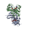
| Title | SUBSTRATE INTERACTIONS BETWEEN TRYPANOTHIONE REDUCTASE AND N1-GLUTATHIONYLSPERMIDINE DISULPHIDE AT 0.28-NM RESOLUTION | ||||||
 Components Components | TRYPANOTHIONE REDUCTASE | ||||||
 Keywords Keywords | OXIDOREDUCTASE | ||||||
| Function / homology |  Function and homology information Function and homology informationtrypanothione-disulfide reductase / trypanothione-disulfide reductase (NADPH) activity / glutathione-disulfide reductase (NADPH) activity / cell redox homeostasis / glutathione metabolic process / flavin adenine dinucleotide binding / cellular response to oxidative stress / mitochondrion / cytosol Similarity search - Function | ||||||
| Biological species |  Crithidia fasciculata (eukaryote) Crithidia fasciculata (eukaryote) | ||||||
| Method |  X-RAY DIFFRACTION / Resolution: 2.8 Å X-RAY DIFFRACTION / Resolution: 2.8 Å | ||||||
 Authors Authors | Bailey, S. / Hunter, W.N. | ||||||
 Citation Citation | Journal: Eur.J.Biochem. / Year: 1993 Title: Substrate interactions between trypanothione reductase and N1-glutathionylspermidine disulphide at 0.28-nm resolution. Authors: Bailey, S. / Smith, K. / Fairlamb, A.H. / Hunter, W.N. #1: Journal: J.Mol.Biol. / Year: 1992Title: Active Site of Trypanothione Reductase: A Target for Rational Drug Design Authors: Hunter, W.N. / Bailey, S. / Habash, J. / Harrop, S.J. / Helliwell, J.R. / Abogye-Kwarteng, T. / Smith, K. / Fairlamb, A.H. #2: Journal: Science / Year: 1992Title: Crystal Structures of Two Viral Peptides in Complex with Murine Mhc Class I H-2KB Authors: Fremont, D.H. / Matsumura, M. / Stura, E.A. / Peterson, P.A. / Wilson, I.A. #3: Journal: J.Mol.Biol. / Year: 1990Title: Initiating a Crystallographic Study of Trypanothione Reductase Authors: Hunter, W.N. / Smith, K. / Derewenda, Z. / Harrop, S.J. / Habash, J. / Islam, M.S. / Helliwell, J.R. / Fairlamb, A.H. #4: Journal: Nature / Year: 1984Title: A Large Increase in Enzyme-Substrate Affinity by Protein Engineering Authors: Wilkinson, A.J. / Fersht, A.R. / Blow, D.M. / Carter, P. / Winter, G. | ||||||
| History |
|
- Structure visualization
| Structure viewer | Molecule: MolmilJmol/JSmol |
|---|
- Downloads & links
-Download
| PDBx/mmCIF format | 1typ.cif.gz | 213.3 KB | Display | PDBx/mmCIF format |
|---|---|---|---|---|
| PDB format | pdb1typ.ent.gz | 168.8 KB | Display | PDB format |
| PDBx/mmJSON format | 1typ.json.gz | Tree view | PDBx/mmJSON format | |
| Others |  Other downloads Other downloads |
-Validation report
| Arichive directory | https://data.pdbj.org/pub/pdb/validation_reports/ty/1typftp://data.pdbj.org/pub/pdb/validation_reports/ty/1typ | HTTPS FTP |
|---|
-Related structure data
| Similar structure data |
|---|

-Links
 PDBj
PDBj


- Assembly
| Deposited unit | 
| ||||||||
|---|---|---|---|---|---|---|---|---|---|
| 1 |
| ||||||||
| Unit cell |
| ||||||||
| Atom site foot note | 1: CIS PROLINE - PRO A 43 / 2: CIS PROLINE - PRO A 370 / 3: CIS PROLINE - PRO A 462 / 4: CIS PROLINE - PRO B 43 / 5: CIS PROLINE - PRO B 370 / 6: CIS PROLINE - PRO B 462 |
-Components
-Protein , 1 types, 2 molecules AB
| #1: Protein | Mass: 52901.805 Da / Num. of mol.: 2 Source method: isolated from a genetically manipulated source Source: (gene. exp.) Crithidia fasciculata (eukaryote) / References: UniProt: P39040, EC: 1.6.4.8 |
|---|
-Non-polymers , 5 types, 412 molecules 


| #2: Chemical |  Mass: 785.550 Da / Num. of mol.: 2 / Source method: obtained synthetically / Formula: C27H33N9O15P2 / Comment: FAD*YM Mass: 785.550 Da / Num. of mol.: 2 / Source method: obtained synthetically / Formula: C27H33N9O15P2 / Comment: FAD*YM#3: Chemical |  Mass: 743.405 Da / Num. of mol.: 2 / Source method: obtained synthetically / Formula: C21H28N7O17P3 Mass: 743.405 Da / Num. of mol.: 2 / Source method: obtained synthetically / Formula: C21H28N7O17P3#4: Chemical | ChemComp-GSH /  Mass: 307.323 Da / Num. of mol.: 4 / Source method: obtained synthetically / Formula: C10H17N3O6S Mass: 307.323 Da / Num. of mol.: 4 / Source method: obtained synthetically / Formula: C10H17N3O6S#5: Chemical | ChemComp-SPD /  Mass: 145.246 Da / Num. of mol.: 4 / Source method: obtained synthetically / Formula: C7H19N3 Mass: 145.246 Da / Num. of mol.: 4 / Source method: obtained synthetically / Formula: C7H19N3#6: Water | ChemComp-HOH / | Mass: 18.015 Da / Num. of mol.: 400 / Source method: isolated from a natural source / Formula: H2O |
|---|
-Details
| Nonpolymer details | N1-GLUTATHION |
|---|
-Experimental details
-Experiment
| Experiment | Method: X-RAY DIFFRACTION |
|---|
- Sample preparation
| Crystal | Density Matthews: 3.62 Å3/Da / Density % sol: 66.07 % | |||||||||||||||||||||||||
|---|---|---|---|---|---|---|---|---|---|---|---|---|---|---|---|---|---|---|---|---|---|---|---|---|---|---|
| Crystal grow | *PLUS Temperature: 4 ℃ / pH: 7 / Method: vapor diffusion, hanging drop | |||||||||||||||||||||||||
| Components of the solutions | *PLUS
|
-Data collection
| Reflection | *PLUS Highest resolution: 2.8 Å / Lowest resolution: 8.85 Å / Num. obs: 35848 / % possible obs: 70 % / Rmerge(I) obs: 0.096 |
|---|---|
| Reflection shell | *PLUS Highest resolution: 2.8 Å / Lowest resolution: 2.95 Å / % possible obs: 36.9 % / Num. possible: 5076 / Rmerge(I) obs: 0.24 |
- Processing
| Software |
| ||||||||||||||||||||||||||||||||||||||||||||||||||||||||||||
|---|---|---|---|---|---|---|---|---|---|---|---|---|---|---|---|---|---|---|---|---|---|---|---|---|---|---|---|---|---|---|---|---|---|---|---|---|---|---|---|---|---|---|---|---|---|---|---|---|---|---|---|---|---|---|---|---|---|---|---|---|---|
| Refinement | Resolution: 2.8→8 Å / σ(F): 1 Details: REFERENCE 1 DESCRIBES THE STRUCTURE SOLUTION BY MOLECULAR REPLACEMENT AND PARTIAL REFINEMENT TO ALLOW DETAILS OF THE ACTIVE SITE TO BE PRESENTED.
| ||||||||||||||||||||||||||||||||||||||||||||||||||||||||||||
| Refinement step | Cycle: LAST / Resolution: 2.8→8 Å
| ||||||||||||||||||||||||||||||||||||||||||||||||||||||||||||
| Refine LS restraints |
| ||||||||||||||||||||||||||||||||||||||||||||||||||||||||||||
| Software | *PLUS Name: X-PLOR / Classification: refinement | ||||||||||||||||||||||||||||||||||||||||||||||||||||||||||||
| Refinement | *PLUS Highest resolution: 2.8 Å / Lowest resolution: 8 Å / Num. reflection obs: 32393 / σ(F): 1 / Rfactor obs: 0.148 | ||||||||||||||||||||||||||||||||||||||||||||||||||||||||||||
| Solvent computation | *PLUS | ||||||||||||||||||||||||||||||||||||||||||||||||||||||||||||
| Displacement parameters | *PLUS | ||||||||||||||||||||||||||||||||||||||||||||||||||||||||||||
| Refine LS restraints | *PLUS Type: x_angle_d / Dev ideal: 3.3 |
