[English] 日本語
 Yorodumi
- PDB-1gxf: CRYSTAL STRUCTURE OF TRYPANOSOMA CRUZI TRYPANOTHIONE REDUCTASE IN... -
+ Open data
- Basic information
| Entry | Database: PDB / ID: 1gxf | ||||||
|---|---|---|---|---|---|---|---|
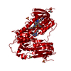
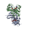
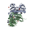
| Title | CRYSTAL STRUCTURE OF TRYPANOSOMA CRUZI TRYPANOTHIONE REDUCTASE IN COMPLEX WITH THE INHIBITOR QUINACRINE MUSTARD | ||||||
 Components Components | TRYPANOTHIONE REDUCTASE (OXIDIZED FORM) | ||||||
 Keywords Keywords | OXIDOREDUCTASE / TRYPANOTHIONE REDUCTASE / FAD DEPENDENT / DISULPHIDE OXIDOREDUCTASE / QUINACRINE MUSTARD / INHIBITOR / REDOX-ACTIVE CENTER / FLAVOPROTEIN / FAD / NADP | ||||||
| Function / homology |  Function and homology information Function and homology informationtrypanothione-disulfide reductase / trypanothione-disulfide reductase (NADPH) activity / glutathione-disulfide reductase (NADPH) activity / glutathione metabolic process / cell redox homeostasis / flavin adenine dinucleotide binding / cellular response to oxidative stress / mitochondrion / cytosol Similarity search - Function | ||||||
| Biological species |  | ||||||
| Method |  X-RAY DIFFRACTION / X-RAY DIFFRACTION /  SYNCHROTRON / SYNCHROTRON /  MOLECULAR REPLACEMENT / Resolution: 2.7 Å MOLECULAR REPLACEMENT / Resolution: 2.7 Å | ||||||
 Authors Authors | Bond, C.S. / Peterson, M.R. / Vickers, T.J. / Fairlamb, A.H. / Hunter, W.N. | ||||||
 Citation Citation |  Journal: J.Biol.Chem. / Year: 2004 Journal: J.Biol.Chem. / Year: 2004Title: Two Interacting Binding Sites for Quinacrine Derivatives in the Active Site of Trypanothione Reductase: A Template for Drug Design Authors: Saravanamuthu, A. / Vickers, T.J. / Bond, C.S. / Peterson, M.R. / Hunter, W.N. / Fairlamb, A.H. | ||||||
| History |
| ||||||
| Remark 700 | SHEET THE SHEET STRUCTURE OF THIS MOLECULE IS BIFURCATED. IN ORDER TO REPRESENT THIS FEATURE IN ... SHEET THE SHEET STRUCTURE OF THIS MOLECULE IS BIFURCATED. IN ORDER TO REPRESENT THIS FEATURE IN THE SHEET RECORDS BELOW, TWO SHEETS ARE DEFINED. |
- Structure visualization
| Structure viewer | Molecule:  Molmil Molmil Jmol/JSmol Jmol/JSmol |
|---|
- Downloads & links
- Download
| PDBx/mmCIF format |  1gxf.cif.gz 1gxf.cif.gz | 203.1 KB | Display |  PDBx/mmCIF format PDBx/mmCIF format |
|---|---|---|---|---|
| PDB format |  pdb1gxf.ent.gz pdb1gxf.ent.gz | 162.1 KB | Display |  PDB format PDB format |
| PDBx/mmJSON format |  1gxf.json.gz 1gxf.json.gz | Tree view |  PDBx/mmJSON format PDBx/mmJSON format | |
| Others |  Other downloads Other downloads |
-Validation report
| Summary document |  1gxf_validation.pdf.gz 1gxf_validation.pdf.gz | 1.9 MB | Display |  wwPDB validaton report wwPDB validaton report |
|---|---|---|---|---|
| Full document |  1gxf_full_validation.pdf.gz 1gxf_full_validation.pdf.gz | 1.9 MB | Display | |
| Data in XML |  1gxf_validation.xml.gz 1gxf_validation.xml.gz | 42.1 KB | Display | |
| Data in CIF |  1gxf_validation.cif.gz 1gxf_validation.cif.gz | 54.8 KB | Display | |
| Arichive directory |  https://data.pdbj.org/pub/pdb/validation_reports/gx/1gxf https://data.pdbj.org/pub/pdb/validation_reports/gx/1gxf ftp://data.pdbj.org/pub/pdb/validation_reports/gx/1gxf ftp://data.pdbj.org/pub/pdb/validation_reports/gx/1gxf | HTTPS FTP |
-Related structure data
| Related structure data |  1tytS S: Starting model for refinement |
|---|---|
| Similar structure data |
- Links
Links
- Assembly
| Deposited unit | 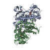
| ||||||||
|---|---|---|---|---|---|---|---|---|---|
| 1 |
| ||||||||
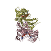
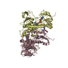
| Unit cell |
| ||||||||
| Noncrystallographic symmetry (NCS) | NCS oper: (Code: given Matrix: (-0.979689, 0.200521, 0.000849), Vector: |
- Components
| #1: Protein | Mass: 53925.668 Da / Num. of mol.: 2 Source method: isolated from a genetically manipulated source Details: COVALENTLY LABELLED WITH QUINACRINE MUSTARD / Source: (gene. exp.)   #2: Chemical | #3: Chemical | ChemComp-MAE / | #4: Chemical | ChemComp-QUM / #5: Water | ChemComp-HOH / | Compound details | TRYPANOTHIONE IS THE PARASITE ANALOG OF GLUTATHIONE AND THIS ENZYME IS THE EQUIVALENT OF ...TRYPANOTHI | Has protein modification | Y | |
|---|
-Experimental details
-Experiment
| Experiment | Method:  X-RAY DIFFRACTION / Number of used crystals: 1 X-RAY DIFFRACTION / Number of used crystals: 1 |
|---|
- Sample preparation
| Crystal | Density Matthews: 3.17 Å3/Da / Density % sol: 61 % |
|---|---|
| Crystal grow | pH: 6 / Details: MALEIC BUFFER, PEG 4000, pH 6.00 |
-Data collection
| Diffraction | Mean temperature: 293 K |
|---|---|
| Diffraction source | Source:  SYNCHROTRON / Site: SYNCHROTRON / Site:  ESRF ESRF  / Beamline: BM14 / Wavelength: 0.92 / Beamline: BM14 / Wavelength: 0.92 |
| Detector | Type: MARRESEARCH / Detector: IMAGE PLATE |
| Radiation | Protocol: SINGLE WAVELENGTH / Monochromatic (M) / Laue (L): M / Scattering type: x-ray |
| Radiation wavelength | Wavelength: 0.92 Å / Relative weight: 1 |
| Reflection | Resolution: 2.7→21 Å / Num. obs: 29073 / % possible obs: 79 % / Observed criterion σ(I): 0 / Redundancy: 3 % / Rmerge(I) obs: 0.08 / Net I/σ(I): 9.4 |
| Reflection shell | Resolution: 2.7→2.9 Å / Rmerge(I) obs: 0.34 / Mean I/σ(I) obs: 2.6 / % possible all: 82 |
- Processing
| Software |
| ||||||||||||||||
|---|---|---|---|---|---|---|---|---|---|---|---|---|---|---|---|---|---|
| Refinement | Method to determine structure:  MOLECULAR REPLACEMENT MOLECULAR REPLACEMENTStarting model: PDB ENTRY 1TYT Resolution: 2.7→21 Å / SU B: 11.848 / SU ML: 0.25126 / Cross valid method: THROUGHOUT / σ(F): 0 / ESU R Free: 0.3924 Details: THE MISSING N- AND C-TERMINAL RESIDUES COULD NOT BE SEEN IN THE ELECTRON DENSITY MAP CLEARLY
| ||||||||||||||||
| Displacement parameters | Biso mean: 46 Å2 | ||||||||||||||||
| Refinement step | Cycle: LAST / Resolution: 2.7→21 Å
|
 Movie
Movie Controller






 PDBj
PDBj


