 Movie
Movie Controller


+ Open data

- Basic information
| Entry | Database: PDB / ID: 1aog | ||||||
|---|---|---|---|---|---|---|---|
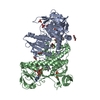
| Title | TRYPANOSOMA CRUZI TRYPANOTHIONE REDUCTASE (OXIDIZED FORM) | ||||||
 Components Components | TRYPANOTHIONE REDUCTASE | ||||||
 Keywords Keywords | OXIDOREDUCTASE / TRYPANOTHIONE REDUCTASE / FAD DEPENDENT DISULPHIDE OXIDOREDUCTASE | ||||||
| Function / homology |  Function and homology information Function and homology informationtrypanothione-disulfide reductase / trypanothione-disulfide reductase (NADPH) activity / glutathione-disulfide reductase (NADPH) activity / cell redox homeostasis / glutathione metabolic process / flavin adenine dinucleotide binding / cellular response to oxidative stress / mitochondrion / cytosol Similarity search - Function | ||||||
| Biological species |  | ||||||
| Method |  X-RAY DIFFRACTION / SYNCHROTRON / MOLECULAR REPLACEMENT / Resolution: 2.3 Å X-RAY DIFFRACTION / SYNCHROTRON / MOLECULAR REPLACEMENT / Resolution: 2.3 Å | ||||||
 Authors Authors | Bond, C.S. / Zhang, Y. / Hunter, W.N. | ||||||
 Citation Citation | Journal: Protein Sci. / Year: 1996 Title: The crystal structure of trypanothione reductase from the human pathogen Trypanosoma cruzi at 2.3 A resolution. Authors: Zhang, Y. / Bond, C.S. / Bailey, S. / Cunningham, M.L. / Fairlamb, A.H. / Hunter, W.N. #1: Journal: Eur.J.Biochem. / Year: 1995Title: Site-Directed Mutagenesis of the Redox-Active Cysteines of Trypanosoma Cruzi Trypanothione Reductase Authors: Borges, A. / Cunningham, M.L. / Tovar, J. / Fairlamb, A.H. #2: Journal: J.Mol.Biol. / Year: 1993Title: Trypanosoma Cruzi Trypanothione Reductase Crystallisation, Unit Cell Dimensions and Structure Solution Authors: Zhang, Y. / Bailey, S. / Naismith, J.H. / Bond, C.S. / Habash, J. / Mclaughlin, P. / Papiz, M.Z. / Borges, A. / Cunningham, M.L. / Fairlamb, A.H. / Hunter, W.N. | ||||||
| History |
|
- Structure visualization
| Structure viewer | Molecule: MolmilJmol/JSmol |
|---|
- Downloads & links
-Download
| PDBx/mmCIF format | 1aog.cif.gz | 209.9 KB | Display | PDBx/mmCIF format |
|---|---|---|---|---|
| PDB format | pdb1aog.ent.gz | 165.3 KB | Display | PDB format |
| PDBx/mmJSON format | 1aog.json.gz | Tree view | PDBx/mmJSON format | |
| Others |  Other downloads Other downloads |
-Validation report
| Arichive directory | https://data.pdbj.org/pub/pdb/validation_reports/ao/1aogftp://data.pdbj.org/pub/pdb/validation_reports/ao/1aog | HTTPS FTP |
|---|
-Related structure data
| Related structure data |  1tytS S: Starting model for refinement |
|---|---|
| Similar structure data |


-Links
 PDBj
PDBj


- Assembly
| Deposited unit | 
| ||||||||
|---|---|---|---|---|---|---|---|---|---|
| 1 |
| ||||||||
| Unit cell |
|
-Components
| #1: Protein | Mass: 53175.773 Da / Num. of mol.: 2 / Source method: isolated from a natural source / Source: (natural) #2: Chemical |   Mass: 785.550 Da / Num. of mol.: 2 / Source method: obtained synthetically / Formula: C27H33N9O15P2 / Comment: FAD*YM Mass: 785.550 Da / Num. of mol.: 2 / Source method: obtained synthetically / Formula: C27H33N9O15P2 / Comment: FAD*YM#3: Chemical |   Mass: 116.072 Da / Num. of mol.: 2 / Source method: obtained synthetically / Formula: C4H4O4 Mass: 116.072 Da / Num. of mol.: 2 / Source method: obtained synthetically / Formula: C4H4O4#4: Water | ChemComp-HOH / |  Mass: 18.015 Da / Num. of mol.: 419 / Source method: isolated from a natural source / Formula: H2O Mass: 18.015 Da / Num. of mol.: 419 / Source method: isolated from a natural source / Formula: H2OHas protein modification | Y | |
|---|
-Experimental details
-Experiment
| Experiment | Method: X-RAY DIFFRACTION / Number of used crystals: 7 |
|---|
- Sample preparation
| Crystal | Density Matthews: 3.17 Å3/Da / Density % sol: 61.21 % | |||||||||||||||||||||||||||||||||||
|---|---|---|---|---|---|---|---|---|---|---|---|---|---|---|---|---|---|---|---|---|---|---|---|---|---|---|---|---|---|---|---|---|---|---|---|---|
| Crystal grow | pH: 6 / Details: pH 6.0 | |||||||||||||||||||||||||||||||||||
| Crystal grow | *PLUS Temperature: 6-8 ℃ / Method: vapor diffusion, hanging drop | |||||||||||||||||||||||||||||||||||
| Components of the solutions | *PLUS
|
-Data collection
| Diffraction | Mean temperature: 293 K |
|---|---|
| Diffraction source | Source: SYNCHROTRON / Site: SRS  / Beamline: PX9.6 / Wavelength: 0.87 / Beamline: PX9.6 / Wavelength: 0.87 |
| Detector | Type: RIGAKU RAXIS / Detector: IMAGE PLATE / Date: Mar 1, 1991 |
| Radiation | Monochromatic (M) / Laue (L): M / Scattering type: x-ray |
| Radiation wavelength | Wavelength: 0.87 Å / Relative weight: 1 |
| Reflection | Resolution: 2.3→20 Å / Num. obs: 55392 / % possible obs: 94.1 % / Observed criterion σ(I): 1 / Redundancy: 3.1 % / Rmerge(I) obs: 0.109 |
| Reflection | *PLUS Num. measured all: 170106 |
| Reflection shell | *PLUS Highest resolution: 2.3 Å / Lowest resolution: 2.4 Å / % possible obs: 80 % |
- Processing
| Software |
| ||||||||||||||||||||||||||||||||||||||||||||||||||||||||||||
|---|---|---|---|---|---|---|---|---|---|---|---|---|---|---|---|---|---|---|---|---|---|---|---|---|---|---|---|---|---|---|---|---|---|---|---|---|---|---|---|---|---|---|---|---|---|---|---|---|---|---|---|---|---|---|---|---|---|---|---|---|---|
| Refinement | Method to determine structure: MOLECULAR REPLACEMENT Starting model: PDB ENTRY 1TYT Resolution: 2.3→8 Å / σ(F): 1
| ||||||||||||||||||||||||||||||||||||||||||||||||||||||||||||
| Displacement parameters | Biso mean: 32.4 Å2 | ||||||||||||||||||||||||||||||||||||||||||||||||||||||||||||
| Refinement step | Cycle: LAST / Resolution: 2.3→8 Å
| ||||||||||||||||||||||||||||||||||||||||||||||||||||||||||||
| Refine LS restraints |
| ||||||||||||||||||||||||||||||||||||||||||||||||||||||||||||
| Software | *PLUS Name: X-PLOR / Version: 3.1 / Classification: refinement | ||||||||||||||||||||||||||||||||||||||||||||||||||||||||||||
| Refinement | *PLUS | ||||||||||||||||||||||||||||||||||||||||||||||||||||||||||||
| Solvent computation | *PLUS | ||||||||||||||||||||||||||||||||||||||||||||||||||||||||||||
| Displacement parameters | *PLUS | ||||||||||||||||||||||||||||||||||||||||||||||||||||||||||||
| Refine LS restraints | *PLUS
|
