 Movie
Movie Controller

[English] 日本語
 Yorodumi
- PDB-3lad: REFINED CRYSTAL STRUCTURE OF LIPOAMIDE DEHYDROGENASE FROM AZOTOBA... -
+ Open data

- Basic information
| Entry | Database: PDB / ID: 3lad | ||||||
|---|---|---|---|---|---|---|---|
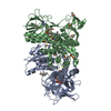
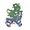
| Title | REFINED CRYSTAL STRUCTURE OF LIPOAMIDE DEHYDROGENASE FROM AZOTOBACTER VINELANDII AT 2.2 ANGSTROMS RESOLUTION. A COMPARISON WITH THE STRUCTURE OF GLUTATHIONE REDUCTASE | ||||||
 Components Components | DIHYDROLIPOAMIDE DEHYDROGENASE | ||||||
 Keywords Keywords | OXIDOREDUCTASE | ||||||
| Function / homology |  Function and homology information Function and homology informationdihydrolipoyl dehydrogenase / dihydrolipoyl dehydrogenase (NADH) activity / 2-oxoglutarate metabolic process / flavin adenine dinucleotide binding / cytoplasm Similarity search - Function | ||||||
| Biological species |  Azotobacter vinelandii (bacteria) Azotobacter vinelandii (bacteria) | ||||||
| Method |  X-RAY DIFFRACTION / Resolution: 2.2 Å X-RAY DIFFRACTION / Resolution: 2.2 Å | ||||||
 Authors Authors | Mattevi, A. / Schierbeek, A.J. / Hol, W.G.J. | ||||||
 Citation Citation | Journal: J.Mol.Biol. / Year: 1991 Title: Refined crystal structure of lipoamide dehydrogenase from Azotobacter vinelandii at 2.2 A resolution. A comparison with the structure of glutathione reductase. Authors: Mattevi, A. / Schierbeek, A.J. / Hol, W.G. #1: Journal: J.Mol.Biol. / Year: 1989Title: X-Ray Structure of Lipoamide Dehydrogenase from Azotobacter Vinelandii Determined by a Combination of Molecular and Isomorphous Replacement Techniques Authors: Schierbeek, A.J. / Swarte, M.B.A. / Dijkstra, B.W. / Vriend, G. / Read, R.J. / Hol, W.G.J. / Drenth, J. / Betzel, C. #2: Journal: Eur.J.Biochem. / Year: 1988Title: Lipoamide Dehydrogenase from Azotobacter Vinelandii: Molecular Cloning, Organization and Sequence Analysis of the Gene Authors: Westphal, A.H. / Dekok, A. #3: Journal: J.Mol.Biol. / Year: 1983Title: Crystallization and Preliminary X-Ray Investigation of Lipoamide Dehydrogenase from Azotobacter Vinelandii Authors: Schierbeek, A.J. / Van Derlaan, J.M. / Groendijk, H. / Wierenga, R.K. / Drenth, J. | ||||||
| History |
|
- Structure visualization
| Structure viewer | Molecule: MolmilJmol/JSmol |
|---|
- Downloads & links
-Download
| PDBx/mmCIF format | 3lad.cif.gz | 193.3 KB | Display | PDBx/mmCIF format |
|---|---|---|---|---|
| PDB format | pdb3lad.ent.gz | 152.4 KB | Display | PDB format |
| PDBx/mmJSON format | 3lad.json.gz | Tree view | PDBx/mmJSON format | |
| Others |  Other downloads Other downloads |
-Validation report
| Arichive directory | https://data.pdbj.org/pub/pdb/validation_reports/la/3ladftp://data.pdbj.org/pub/pdb/validation_reports/la/3lad | HTTPS FTP |
|---|
-Related structure data
| Similar structure data |
|---|


-Links
 PDBj
PDBj


- Assembly
| Deposited unit | 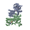
| ||||||||
|---|---|---|---|---|---|---|---|---|---|
| 1 |
| ||||||||
| Unit cell |
| ||||||||
| Atom site foot note | 1: HIS A 359 - PRO A 360 OMEGA =122.43 PEPTIDE BOND DEVIATES SIGNIFICANTLY FROM TRANS CONFORMATION 2: CIS PROLINE - PRO A 451 3: HIS B 359 - PRO B 360 OMEGA =324.57 PEPTIDE BOND DEVIATES SIGNIFICANTLY FROM TRANS CONFORMATION 4: CIS PROLINE - PRO B 451 | ||||||||
| Noncrystallographic symmetry (NCS) | NCS oper: (Code: given Matrix: (0.083696, 0.801095, 0.592657), Vector: Details | THE TRANSFORMATION PROVIDED ON *MTRIX* RECORDS BELOW WILL YIELD APPROXIMATE COORDINATES FOR CHAIN *A* WHEN APPLIED TO CHAIN *B*. AFTER SUPERPOSITION, THE RMS DIFFERENCE BETWEEN THE POSITIONS OF 472 EQUIVALENT CARBON ALPHA ATOMS IS 0.63 ANGSTROMS. | |
-Components
| #1: Protein | Mass: 49494.750 Da / Num. of mol.: 2 Source method: isolated from a genetically manipulated source Source: (gene. exp.) Azotobacter vinelandii (bacteria) / References: UniProt: P18925, dihydrolipoyl dehydrogenase#2: Chemical |   Mass: 785.550 Da / Num. of mol.: 2 / Source method: obtained synthetically / Formula: C27H33N9O15P2 / Comment: FAD*YM Mass: 785.550 Da / Num. of mol.: 2 / Source method: obtained synthetically / Formula: C27H33N9O15P2 / Comment: FAD*YM#3: Water | ChemComp-HOH / |  Mass: 18.015 Da / Num. of mol.: 451 / Source method: isolated from a natural source / Formula: H2O Mass: 18.015 Da / Num. of mol.: 451 / Source method: isolated from a natural source / Formula: H2OHas protein modification | Y | |
|---|
-Experimental details
-Experiment
| Experiment | Method: X-RAY DIFFRACTION |
|---|
- Sample preparation
| Crystal | Density Matthews: 2.61 Å3/Da / Density % sol: 52.81 % | |||||||||||||||
|---|---|---|---|---|---|---|---|---|---|---|---|---|---|---|---|---|
| Crystal grow | *PLUS Temperature: 20 ℃ / pH: 7.5 / Method: vapor diffusion, hanging drop | |||||||||||||||
| Components of the solutions | *PLUS
|
- Processing
| Software |
| ||||||||||||||||||||||||||||||||||||||||||||||||||||||||||||
|---|---|---|---|---|---|---|---|---|---|---|---|---|---|---|---|---|---|---|---|---|---|---|---|---|---|---|---|---|---|---|---|---|---|---|---|---|---|---|---|---|---|---|---|---|---|---|---|---|---|---|---|---|---|---|---|---|---|---|---|---|---|
| Refinement | Highest resolution: 2.2 Å / σ(F): 2 /
| ||||||||||||||||||||||||||||||||||||||||||||||||||||||||||||
| Refinement step | Cycle: LAST / Highest resolution: 2.2 Å
| ||||||||||||||||||||||||||||||||||||||||||||||||||||||||||||
| Refine LS restraints |
| ||||||||||||||||||||||||||||||||||||||||||||||||||||||||||||
| Refinement | *PLUS Highest resolution: 2.2 Å / Lowest resolution: 8 Å / Num. reflection obs: 44294 / σ(F): 2 / Rfactor obs: 0.192 | ||||||||||||||||||||||||||||||||||||||||||||||||||||||||||||
| Solvent computation | *PLUS | ||||||||||||||||||||||||||||||||||||||||||||||||||||||||||||
| Displacement parameters | *PLUS |
