Journal: To Be Published Title: {Ru(CO)x}-core Complexes with N-Methylbenzimidazole (MBI) and 5,6-Dimethylbenzimidazole (DMBI) as Possible Metal Based Drugs and CO Release Materials. Synthesis, X-ray Structure and Evaluation ...Title: {Ru(CO)x}-core Complexes with N-Methylbenzimidazole (MBI) and 5,6-Dimethylbenzimidazole (DMBI) as Possible Metal Based Drugs and CO Release Materials. Synthesis, X-ray Structure and Evaluation of Cytotoxic Activity against Human Cancer Cells Authors: Merlino, A.
Mass: 18.015 Da / Num. of mol.: 159 / Source method: isolated from a natural source / Formula: H2O
-
Details
Has protein modification
Y
-
Experimental details
-
Experiment
Experiment
Method: X-RAY DIFFRACTION
-
Sample preparation
Crystal
Density Matthews: 1.95 Å3/Da / Density % sol: 36.97 %
Crystal grow
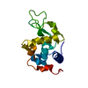
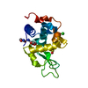
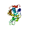
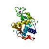
Temperature: 298 K / Method: vapor diffusion, hanging drop Details: Hen egg white lysozyme was incubated for 2 h in the presence of Ru-MBI in 1:10 protein to metal compound ratio. The adduct was then crystallized by using the hanging drop vapour diffusion ...Details: Hen egg white lysozyme was incubated for 2 h in the presence of Ru-MBI in 1:10 protein to metal compound ratio. The adduct was then crystallized by using the hanging drop vapour diffusion method. Drops of 1 microliter were prepared at room temperature mixing 0.5 microliter of adduct at concentration 15 mg/mL with an equal amount of a reservoir constituted by 1.1 M NaCl, 0.1 M acetate buffer, pH 4.0. Crystals of the adduct grow within 24-48 hours. PH range: 4
Protocol: SINGLE WAVELENGTH / Monochromatic (M) / Laue (L): M / Scattering type: x-ray
Radiation wavelength
Wavelength: 1.5418 Å / Relative weight: 1
Reflection
Resolution: 2.25→55.5 Å / Num. obs: 5740 / % possible obs: 99.4 % / Redundancy: 6.2 % / Rmerge(I) obs: 0.085 / Net I/σ(I): 12.9
Reflection shell
Resolution: 2.25→2.29 Å / Redundancy: 6.3 % / Rmerge(I) obs: 0.436 / Mean I/σ(I) obs: 5 / % possible all: 100
-
Processing
Software
Name
Version
Classification
REFMAC
5.8.0049
refinement
HKL-2000
datareduction
HKL-2000
datascaling
PHASER
phasing
Refinement
Resolution: 2.25→55.5 Å / Cor.coef. Fo:Fc: 0.966 / Cor.coef. Fo:Fc free: 0.929 / SU B: 7.658 / SU ML: 0.185 / Cross valid method: THROUGHOUT / ESU R: 0.456 / ESU R Free: 0.254 / Stereochemistry target values: MAXIMUM LIKELIHOOD / Details: HYDROGENS HAVE BEEN ADDED IN THE RIDING POSITIONS
Rfactor
Num. reflection
% reflection
Selection details
Rfree
0.23734
261
4.6 %
RANDOM
Rwork
0.15228
-
-
-
obs
0.15626
5452
99.41 %
-
Solvent computation
Ion probe radii: 0.8 Å / Shrinkage radii: 0.8 Å / VDW probe radii: 1.2 Å / Solvent model: MASK
 Movie
Movie Controller

 Yorodumi
 Open data
 Basic information
 Components
 Keywords
 Function and homology information
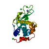
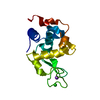
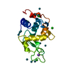
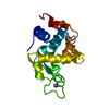
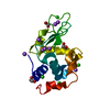
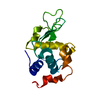
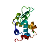
 X-RAY DIFFRACTION / Resolution: 2.25 Å
  Authors
 Citation
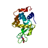
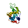
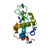
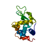
 Structure visualization
 Downloads & links
 Other downloads
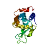


 PDBj
PDBj


 Assembly
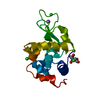


 Mass: 232.606 Da / Num. of mol.: 1 / Source method: obtained synthetically / Formula: C2H8ClO4Ru
 Mass: 28.010 Da / Num. of mol.: 1 / Source method: obtained synthetically / Formula: CO
 Mass: 22.990 Da / Num. of mol.: 1 / Source method: obtained synthetically / Formula: Na
 Mass: 35.453 Da / Num. of mol.: 3 / Source method: obtained synthetically / Formula: Cl
 Sample preparation
 Processing