 Movie
Movie Controller

[English] 日本語
 Yorodumi
- PDB-3hfm: STRUCTURE OF AN ANTIBODY-ANTIGEN COMPLEX. CRYSTAL STRUCTURE OF TH... -
+ Open data

- Basic information
| Entry | Database: PDB / ID: 3hfm | ||||||
|---|---|---|---|---|---|---|---|
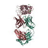
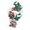
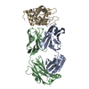
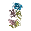
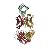
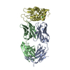
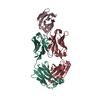
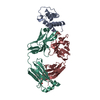
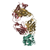
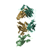
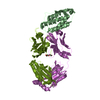
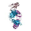
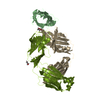
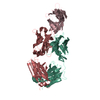
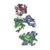
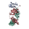
| Title | STRUCTURE OF AN ANTIBODY-ANTIGEN COMPLEX. CRYSTAL STRUCTURE OF THE HY/HEL-10 FAB-LYSOZYME COMPLEX | ||||||
 Components Components |
| ||||||
 Keywords Keywords | COMPLEX(ANTIBODY-ANTIGEN) | ||||||
| Function / homology |  Function and homology information Function and homology informationLactose synthesis / Antimicrobial peptides / Neutrophil degranulation / beta-N-acetylglucosaminidase activity / cell wall macromolecule catabolic process / lysozyme / lysozyme activity / killing of cells of another organism / defense response to Gram-negative bacterium / defense response to bacterium ...Lactose synthesis / Antimicrobial peptides / Neutrophil degranulation / beta-N-acetylglucosaminidase activity / cell wall macromolecule catabolic process / lysozyme / lysozyme activity / killing of cells of another organism / defense response to Gram-negative bacterium / defense response to bacterium / defense response to Gram-positive bacterium / endoplasmic reticulum / : / identical protein binding / cytoplasm Similarity search - Function | ||||||
| Biological species |   | ||||||
| Method |  X-RAY DIFFRACTION / Resolution: 3 Å X-RAY DIFFRACTION / Resolution: 3 Å | ||||||
 Authors Authors | Padlan, E.A. / Davies, D.R. | ||||||
 Citation Citation | Journal: Proc.Natl.Acad.Sci.USA / Year: 1989 Title: Structure of an antibody-antigen complex: crystal structure of the HyHEL-10 Fab-lysozyme complex. Authors: Padlan, E.A. / Silverton, E.W. / Sheriff, S. / Cohen, G.H. / Smith-Gill, S.J. / Davies, D.R. #1: Journal: J.Mol.Biol. / Year: 1987Title: A Three-Dimensional Model of an Anti-Lysozyme Antibody Authors: Smith-Gill, S.J. / Mainhart, C. / Lavoie, T.B. / Feldmann, R.J. / Drohan, W. / Brooks, B.R. #2: Journal: J.Mol.Biol. / Year: 1984Title: Crystalline Monoclonal Antibody Fabs Complexed to Hen Egg White Lysozyme Authors: Silverton, E.W. / Padlan, E.A. / Davies, D.R. / Smith-Gill, S. / Potter, M. | ||||||
| History |
|
- Structure visualization
| Structure viewer | Molecule: MolmilJmol/JSmol |
|---|
- Downloads & links
-Download
| PDBx/mmCIF format | 3hfm.cif.gz | 115.5 KB | Display | PDBx/mmCIF format |
|---|---|---|---|---|
| PDB format | pdb3hfm.ent.gz | 88.8 KB | Display | PDB format |
| PDBx/mmJSON format | 3hfm.json.gz | Tree view | PDBx/mmJSON format | |
| Others |  Other downloads Other downloads |
-Validation report
| Arichive directory | https://data.pdbj.org/pub/pdb/validation_reports/hf/3hfmftp://data.pdbj.org/pub/pdb/validation_reports/hf/3hfm | HTTPS FTP |
|---|
-Related structure data
| Similar structure data |
|---|

-Links
 PDBj
PDBj


- Assembly
| Deposited unit | 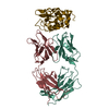
| ||||||||
|---|---|---|---|---|---|---|---|---|---|
| 1 |
| ||||||||
| Unit cell |
| ||||||||
| Atom site foot note | 1: RESIDUES PRO L 8, PRO L 95, PRO L 141, PRO H 147, PRO H 149 AND PRO H 187 ARE CIS-PROLINES. |
-Components
| #1: Antibody | Mass: 23552.857 Da / Num. of mol.: 1 Source method: isolated from a genetically manipulated source Source: (gene. exp.) |
|---|---|
| #2: Antibody | Mass: 23322.873 Da / Num. of mol.: 1 Source method: isolated from a genetically manipulated source Source: (gene. exp.) |
| #3: Protein | Mass: 14331.160 Da / Num. of mol.: 1 Source method: isolated from a genetically manipulated source Source: (gene. exp.) |
| #4: Water | ChemComp-HOH /  Mass: 18.015 Da / Num. of mol.: 1 / Source method: isolated from a natural source / Formula: H2O Mass: 18.015 Da / Num. of mol.: 1 / Source method: isolated from a natural source / Formula: H2O |
| Has protein modification | Y |
-Experimental details
-Experiment
| Experiment | Method: X-RAY DIFFRACTION |
|---|
- Sample preparation
| Crystal | Density Matthews: 3.84 Å3/Da / Density % sol: 67.93 % | ||||||||||||||||||||||||||||||
|---|---|---|---|---|---|---|---|---|---|---|---|---|---|---|---|---|---|---|---|---|---|---|---|---|---|---|---|---|---|---|---|
| Crystal grow | *PLUS Method: vapor diffusion, hanging drop / Details: referred to J.Mol.Biol. 180.761-765 1984 | ||||||||||||||||||||||||||||||
| Components of the solutions | *PLUS
|
-Data collection
| Reflection | *PLUS Highest resolution: 3 Å / Lowest resolution: 10 Å / Num. obs: 12501 / % possible obs: 78 % / Observed criterion σ(F): 3 / Rmerge(I) obs: 0.066 |
|---|
- Processing
| Software | Name: PROLSQ / Classification: refinement | |||||||||||||||||||||||||||||||||||||||||||||||||||||||||||||||
|---|---|---|---|---|---|---|---|---|---|---|---|---|---|---|---|---|---|---|---|---|---|---|---|---|---|---|---|---|---|---|---|---|---|---|---|---|---|---|---|---|---|---|---|---|---|---|---|---|---|---|---|---|---|---|---|---|---|---|---|---|---|---|---|---|
| Refinement | Resolution: 3→10 Å / Rfactor obs: 0.246 | |||||||||||||||||||||||||||||||||||||||||||||||||||||||||||||||
| Refinement step | Cycle: LAST / Resolution: 3→10 Å
| |||||||||||||||||||||||||||||||||||||||||||||||||||||||||||||||
| Refine LS restraints |
| |||||||||||||||||||||||||||||||||||||||||||||||||||||||||||||||
| Refinement | *PLUS Rfactor obs: 0.24 / Highest resolution: 3 Å / Lowest resolution: 10 Å | |||||||||||||||||||||||||||||||||||||||||||||||||||||||||||||||
| Solvent computation | *PLUS | |||||||||||||||||||||||||||||||||||||||||||||||||||||||||||||||
| Displacement parameters | *PLUS | |||||||||||||||||||||||||||||||||||||||||||||||||||||||||||||||
| Refine LS restraints | *PLUS
|
