 Movie
Movie Controller

[English] 日本語
 Yorodumi
- PDB-1jmc: SINGLE STRANDED DNA-BINDING DOMAIN OF HUMAN REPLICATION PROTEIN A... -
+ Open data

- Basic information
| Entry | Database: PDB / ID: 1jmc | ||||||
|---|---|---|---|---|---|---|---|
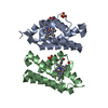
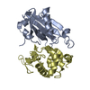
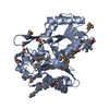
| Title | SINGLE STRANDED DNA-BINDING DOMAIN OF HUMAN REPLICATION PROTEIN A BOUND TO SINGLE STRANDED DNA, RPA70 SUBUNIT, RESIDUES 183-420 | ||||||
 Components Components |
| ||||||
 Keywords Keywords | REPLICATION/DNA / HUMAN SSDNA BINDING REPLICATION PROTEIN A(RPA) / SINGLE STRANDED DNA-BINDING PROTEIN / PROTEIN-SSDNA COMPLEX / COMPLEX (DNA-BINDING PROTEIN-DNA) / REPLICATION-DNA COMPLEX | ||||||
| Function / homology |  Function and homology information Function and homology informationprotein localization to chromosome / DNA replication factor A complex / single-stranded telomeric DNA binding / Removal of the Flap Intermediate / lateral element / G-rich strand telomeric DNA binding / protein localization to site of double-strand break / Mismatch repair (MMR) directed by MSH2:MSH3 (MutSbeta) / Mismatch repair (MMR) directed by MSH2:MSH6 (MutSalpha) / chromatin-protein adaptor activity ...protein localization to chromosome / DNA replication factor A complex / single-stranded telomeric DNA binding / Removal of the Flap Intermediate / lateral element / G-rich strand telomeric DNA binding / protein localization to site of double-strand break / Mismatch repair (MMR) directed by MSH2:MSH3 (MutSbeta) / Mismatch repair (MMR) directed by MSH2:MSH6 (MutSalpha) / chromatin-protein adaptor activity / Removal of the Flap Intermediate from the C-strand / HDR through Single Strand Annealing (SSA) / Impaired BRCA2 binding to RAD51 / hemopoiesis / PCNA-Dependent Long Patch Base Excision Repair / Activation of the pre-replicative complex / Regulation of HSF1-mediated heat shock response / Presynaptic phase of homologous DNA pairing and strand exchange / HSF1 activation / Activation of ATR in response to replication stress / telomere maintenance via telomerase / mismatch repair / SUMOylation of DNA damage response and repair proteins / homeostasis of number of cells within a tissue / site of DNA damage / telomere maintenance / male germ cell nucleus / Translesion synthesis by REV1 / Translesion synthesis by POLK / Translesion synthesis by POLI / Gap-filling DNA repair synthesis and ligation in GG-NER / nucleotide-excision repair / Fanconi Anemia Pathway / Termination of translesion DNA synthesis / meiotic cell cycle / Translesion Synthesis by POLH / base-excision repair / PML body / Recognition of DNA damage by PCNA-containing replication complex / G2/M DNA damage checkpoint / double-strand break repair via homologous recombination / DNA-templated DNA replication / Meiotic recombination / HDR through Homologous Recombination (HRR) / Dual Incision in GG-NER / Formation of Incision Complex in GG-NER / Dual incision in TC-NER / Gap-filling DNA repair synthesis and ligation in TC-NER / single-stranded DNA binding / site of double-strand break / in utero embryonic development / Processing of DNA double-strand break ends / DNA recombination / Regulation of TP53 Activity through Phosphorylation / damaged DNA binding / chromosome, telomeric region / DNA replication / DNA repair / chromatin binding / positive regulation of cell population proliferation / DNA damage response / zinc ion binding / nucleoplasm / nucleus Similarity search - Function | ||||||
| Biological species |  Homo sapiens (human) Homo sapiens (human) | ||||||
| Method |  X-RAY DIFFRACTION / MIRAS / Resolution: 2.4 Å X-RAY DIFFRACTION / MIRAS / Resolution: 2.4 Å | ||||||
 Authors Authors | Bochkarev, A. / Pfuetzner, R. / Edwards, A. / Frappier, L. | ||||||
 Citation Citation | Journal: Nature / Year: 1997 Title: Structure of the single-stranded-DNA-binding domain of replication protein A bound to DNA. Authors: Bochkarev, A. / Pfuetzner, R.A. / Edwards, A.M. / Frappier, L. #1: Journal: J.Biol.Chem. / Year: 1997Title: Replication Protein A. Characterization and Crystallization of the DNA Binding Domain Authors: Pfuetzner, R.A. / Bochkarev, A. / Frappier, L. / Edwards, A.M. | ||||||
| History |
|
- Structure visualization
| Structure viewer | Molecule: MolmilJmol/JSmol |
|---|
- Downloads & links
-Download
| PDBx/mmCIF format | 1jmc.cif.gz | 78.1 KB | Display | PDBx/mmCIF format |
|---|---|---|---|---|
| PDB format | pdb1jmc.ent.gz | 57.8 KB | Display | PDB format |
| PDBx/mmJSON format | 1jmc.json.gz | Tree view | PDBx/mmJSON format | |
| Others |  Other downloads Other downloads |
-Validation report
| Arichive directory | https://data.pdbj.org/pub/pdb/validation_reports/jm/1jmcftp://data.pdbj.org/pub/pdb/validation_reports/jm/1jmc | HTTPS FTP |
|---|
-Related structure data
| Similar structure data |
|---|


-Links
 PDBj
PDBj


- Assembly
| Deposited unit | 
| ||||||||
|---|---|---|---|---|---|---|---|---|---|
| 1 |
| ||||||||
| Unit cell |
|
-Components
| #1: DNA chain | Mass: 2268.497 Da / Num. of mol.: 1 / Source method: obtained synthetically |
|---|---|
| #2: Protein | Mass: 27516.953 Da / Num. of mol.: 1 Source method: isolated from a genetically manipulated source Source: (gene. exp.) Homo sapiens (human) / Plasmid: PET15B / Production host:  |
| #3: Water | ChemComp-HOH /  Mass: 18.015 Da / Num. of mol.: 90 / Source method: isolated from a natural source / Formula: H2O Mass: 18.015 Da / Num. of mol.: 90 / Source method: isolated from a natural source / Formula: H2O |
| Has protein modification | Y |
-Experimental details
-Experiment
| Experiment | Method: X-RAY DIFFRACTION / Number of used crystals: 1 |
|---|
- Sample preparation
| Crystal | Density Matthews: 2.13 Å3/Da / Density % sol: 40 % Description: THREE DIFFERENT MONO-IODINATED AND ONE DI-IODINATED DNA DERIVATIVES WERE USED. | ||||||||||||||||||||||||||||||||||||||||||
|---|---|---|---|---|---|---|---|---|---|---|---|---|---|---|---|---|---|---|---|---|---|---|---|---|---|---|---|---|---|---|---|---|---|---|---|---|---|---|---|---|---|---|---|
| Crystal grow | Method: vapor diffusion, sitting drop / pH: 6.8 Details: 24-27% PEG2000 MME, 20MM NA_CL, 20MM MGCL2, 5MM DTT, 100MM MES PH 6.8, VAPOR DIFFUSION, SITTING DROP | ||||||||||||||||||||||||||||||||||||||||||
| Components of the solutions |
| ||||||||||||||||||||||||||||||||||||||||||
| Crystal | *PLUS Density % sol: 40 % | ||||||||||||||||||||||||||||||||||||||||||
| Crystal grow | *PLUS Temperature: 4 ℃ | ||||||||||||||||||||||||||||||||||||||||||
| Components of the solutions | *PLUS
|
-Data collection
| Diffraction | Mean temperature: 100 K |
|---|---|
| Diffraction source | Source: ROTATING ANODE / Type: RIGAKU RU200 |
| Detector | Type: RIGAKU RAXIS IIC / Detector: IMAGE PLATE / Date: Jun 25, 1996 / Details: MSC |
| Radiation | Protocol: SINGLE WAVELENGTH / Monochromatic (M) / Laue (L): M / Scattering type: x-ray |
| Radiation wavelength | Relative weight: 1 |
| Reflection | Resolution: 2.2→35 Å / Num. obs: 13045 / % possible obs: 95 % / Observed criterion σ(F): 1 / Observed criterion σ(I): 1 / Redundancy: 10.8 % / Rmerge(I) obs: 0.063 |
| Reflection shell | Resolution: 2.2→2.34 Å / Rmerge(I) obs: 0.165 / % possible all: 87.8 |
| Reflection | *PLUS Highest resolution: 2.2 Å / Lowest resolution: 35 Å / % possible obs: 95 % / Num. measured all: 141185 |
| Reflection shell | *PLUS % possible obs: 87.8 % |
- Processing
| Software |
| ||||||||||||||||||||||||||||||||||||||||||||||||||||||||||||||||||||||||||||||||
|---|---|---|---|---|---|---|---|---|---|---|---|---|---|---|---|---|---|---|---|---|---|---|---|---|---|---|---|---|---|---|---|---|---|---|---|---|---|---|---|---|---|---|---|---|---|---|---|---|---|---|---|---|---|---|---|---|---|---|---|---|---|---|---|---|---|---|---|---|---|---|---|---|---|---|---|---|---|---|---|---|---|
| Refinement | Method to determine structure: MIRAS / Resolution: 2.4→6 Å / Data cutoff high absF: 100000 / Data cutoff low absF: 0.1 / Isotropic thermal model: RESTRAINED INDIVIDUAL B F / σ(F): 2 Details: AMINO ACIDS 239 AND 259 ARE IN DISALLOWED REGIONS OF THE RAMACHANDRAN PLOT.
| ||||||||||||||||||||||||||||||||||||||||||||||||||||||||||||||||||||||||||||||||
| Refinement step | Cycle: LAST / Resolution: 2.4→6 Å
| ||||||||||||||||||||||||||||||||||||||||||||||||||||||||||||||||||||||||||||||||
| Refine LS restraints |
| ||||||||||||||||||||||||||||||||||||||||||||||||||||||||||||||||||||||||||||||||
| LS refinement shell | Resolution: 2.4→2.5 Å / Total num. of bins used: 8
| ||||||||||||||||||||||||||||||||||||||||||||||||||||||||||||||||||||||||||||||||
| Software | *PLUS Name: X-PLOR / Version: 3 / Classification: refinement | ||||||||||||||||||||||||||||||||||||||||||||||||||||||||||||||||||||||||||||||||
| Refinement | *PLUS Highest resolution: 2.4 Å / Lowest resolution: 6 Å / σ(F): 2 / % reflection Rfree: 10 % / Rfactor Rfree: 0.33 / Rfactor Rwork: 0.212 | ||||||||||||||||||||||||||||||||||||||||||||||||||||||||||||||||||||||||||||||||
| Solvent computation | *PLUS | ||||||||||||||||||||||||||||||||||||||||||||||||||||||||||||||||||||||||||||||||
| Displacement parameters | *PLUS | ||||||||||||||||||||||||||||||||||||||||||||||||||||||||||||||||||||||||||||||||
| Refine LS restraints | *PLUS
| ||||||||||||||||||||||||||||||||||||||||||||||||||||||||||||||||||||||||||||||||
| LS refinement shell | *PLUS Highest resolution: 2.4 Å / Lowest resolution: 2.5 Å |
