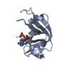
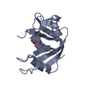
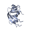
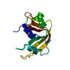
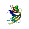
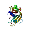
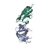
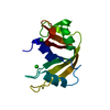
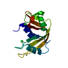
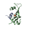
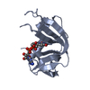
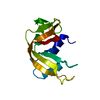
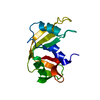
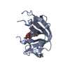
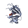
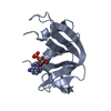
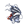
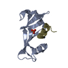
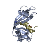
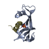
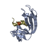
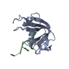
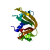
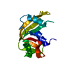
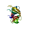
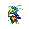
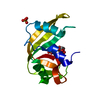
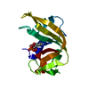
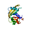
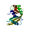
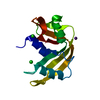
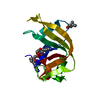
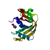
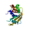
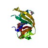
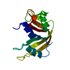
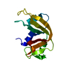
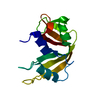
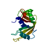
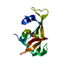
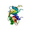
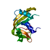
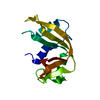
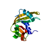
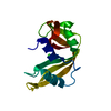
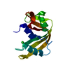
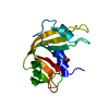
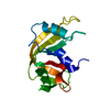
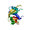
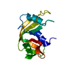
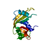
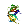
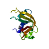
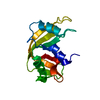
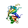
SHEET THE SHEET STRUCTURE OF THIS MOLECULE IS BIFURCATED. IN ORDER TO REPRESENT THIS FEATURE IN ... SHEET THE SHEET STRUCTURE OF THIS MOLECULE IS BIFURCATED. IN ORDER TO REPRESENT THIS FEATURE IN THE SHEET RECORDS BELOW, TWO SHEETS ARE DEFINED.
Resolution: 1.7→73.52 Å / Cor.coef. Fo:Fc: 0.954 / Cor.coef. Fo:Fc free: 0.946 / SU B: 2.116 / SU ML: 0.072 / Cross valid method: THROUGHOUT / ESU R: 0.121 / ESU R Free: 0.116 / Stereochemistry target values: MAXIMUM LIKELIHOOD Details: HYDROGENS HAVE BEEN ADDED IN THE RIDING POSITIONS. DISORDERED SIDE CHAIN ATOMS HAVE BEEN OMITTED. RESIDUES 16-24 AND 87-90 OF EACH CHAIN HAVE POOR DENSITY.
Rfactor
Num. reflection
% reflection
Selection details
Rfree
0.23
1321
5 %
RANDOM
Rwork
0.196
-
-
-
obs
0.198
25134
96.1 %
-
Solvent computation
Ion probe radii: 0.8 Å / Shrinkage radii: 0.8 Å / VDW probe radii: 1.2 Å / Solvent model: MASK
 Movie
Movie Controller


 Open data
 Basic information
 Components
 Keywords
 Function and homology information
 X-RAY DIFFRACTION /
  Authors
 Citation
 Structure visualization
 Downloads & links
 Other downloads
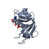


 PDBj
PDBj

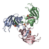
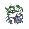
 Assembly
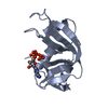
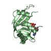

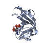
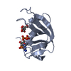
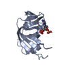
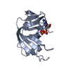
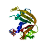
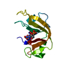
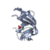
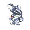
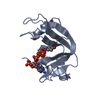
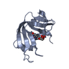
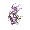
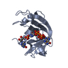
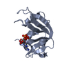
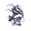
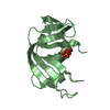
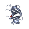
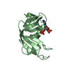
 Mass: 507.181 Da / Num. of mol.: 2 / Source method: obtained synthetically / Formula: C10H16N5O13P3 / Comment: ATP, energy-carrying molecule*YM
 Mass: 18.015 Da / Num. of mol.: 117 / Source method: isolated from a natural source / Formula: H2O
 Sample preparation
 / Beamline: PX14.1 / Wavelength: 0.98
  Processing