 Movie
Movie Controller


+ Open data

- Basic information
| Entry | Database: PDB / ID: 3rsd | ||||||
|---|---|---|---|---|---|---|---|
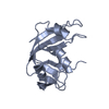
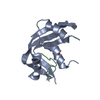
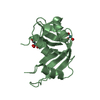
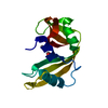
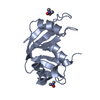
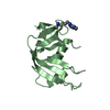
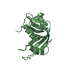
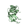
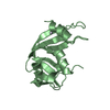
| Title | STRUCTURE OF THE D121N VARIANT OF RIBONUCLEASE A | ||||||
 Components Components | RIBONUCLEASE A | ||||||
 Keywords Keywords | HYDROLASE / ENDONUCLEASE / RIBONUCLEASE A / SITE-DIRECTED MUTAGENESIS | ||||||
| Function / homology |  Function and homology information Function and homology informationpancreatic ribonuclease / ribonuclease A activity / RNA nuclease activity / nucleic acid binding / defense response to Gram-positive bacterium / hydrolase activity / extracellular region Similarity search - Function | ||||||
| Biological species |  | ||||||
| Method |  X-RAY DIFFRACTION / MOLECULAR REPLACEMENT / Resolution: 1.6 Å X-RAY DIFFRACTION / MOLECULAR REPLACEMENT / Resolution: 1.6 Å | ||||||
 Authors Authors | Schultz, L.W. / Quirk, D.J. / Raines, R.T. | ||||||
 Citation Citation | Journal: Biochemistry / Year: 1998 Title: His...Asp catalytic dyad of ribonuclease A: structure and function of the wild-type, D121N, and D121A enzymes. Authors: Schultz, L.W. / Quirk, D.J. / Raines, R.T. | ||||||
| History |
|
- Structure visualization
| Structure viewer | Molecule: MolmilJmol/JSmol |
|---|
- Downloads & links
-Download
| PDBx/mmCIF format | 3rsd.cif.gz | 38.3 KB | Display | PDBx/mmCIF format |
|---|---|---|---|---|
| PDB format | pdb3rsd.ent.gz | 25.8 KB | Display | PDB format |
| PDBx/mmJSON format | 3rsd.json.gz | Tree view | PDBx/mmJSON format | |
| Others |  Other downloads Other downloads |
-Validation report
| Arichive directory | https://data.pdbj.org/pub/pdb/validation_reports/rs/3rsdftp://data.pdbj.org/pub/pdb/validation_reports/rs/3rsd | HTTPS FTP |
|---|
-Related structure data
| Related structure data | 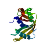 4rsdC 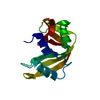 1rphS S: Starting model for refinement C: citing same article ( |
|---|---|
| Similar structure data |

-Links
 PDBj
PDBj

- Assembly
| Deposited unit | 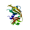
| ||||||||
|---|---|---|---|---|---|---|---|---|---|
| 1 |
| ||||||||
| Unit cell |
|
-Components
| #1: Protein | Mass: 13707.342 Da / Num. of mol.: 1 / Mutation: D121N Source method: isolated from a genetically manipulated source Source: (gene. exp.)  |
|---|---|
| #2: Water | ChemComp-HOH /  Mass: 18.015 Da / Num. of mol.: 100 / Source method: isolated from a natural source / Formula: H2O Mass: 18.015 Da / Num. of mol.: 100 / Source method: isolated from a natural source / Formula: H2O |
| Has protein modification | Y |
-Experimental details
-Experiment
| Experiment | Method: X-RAY DIFFRACTION / Number of used crystals: 1 |
|---|
- Sample preparation
| Crystal | Density Matthews: 2.19 Å3/Da / Density % sol: 60 % | ||||||||||||||||||||
|---|---|---|---|---|---|---|---|---|---|---|---|---|---|---|---|---|---|---|---|---|---|
| Crystal grow | Method: batch method / pH: 5.2 Details: LYOPHILIZED D121N RNASE A WAS DISSOLVED IN UNBUFFERED WATER TO A CONCENTRATION OF 60MG/ ML. CRYSTALLIZATION WAS PERFORMED IN SMALL SILICONIZED TUBES USING THE BATCH METHOD. A SOLUTION (25UL) ...Details: LYOPHILIZED D121N RNASE A WAS DISSOLVED IN UNBUFFERED WATER TO A CONCENTRATION OF 60MG/ ML. CRYSTALLIZATION WAS PERFORMED IN SMALL SILICONIZED TUBES USING THE BATCH METHOD. A SOLUTION (25UL) OF THE ENZYME WAS MIXED WITH AN EQUAL VOLUME OF 95% (V/V) 2-METHYL-2,4-PENTANEDIOL IN 0.10 M MES BUFFER, PH 5.2. CRYSTALS APPEARED WITHIN SEVERAL MONTHS., batch method | ||||||||||||||||||||
| Crystal | *PLUS | ||||||||||||||||||||
| Crystal grow | *PLUS Temperature: 20 ℃ / Method: batch method | ||||||||||||||||||||
| Components of the solutions | *PLUS
|
-Data collection
| Diffraction | Mean temperature: 277 K |
|---|---|
| Diffraction source | Source: ROTATING ANODE / Type: RIGAKU RUH2R / Wavelength: 1.5418 |
| Detector | Type: SIEMENS / Detector: AREA DETECTOR / Date: Aug 28, 1995 / Details: LONG MIRRORS |
| Radiation | Monochromator: NI FILTER / Monochromatic (M) / Laue (L): M / Scattering type: x-ray |
| Radiation wavelength | Wavelength: 1.5418 Å / Relative weight: 1 |
| Reflection | Resolution: 1.6→30 Å / Num. obs: 15756 / % possible obs: 88 % / Observed criterion σ(I): 0.33 / Redundancy: 1.8 % / Rsym value: 0.019 / Net I/σ(I): 31 |
| Reflection shell | Resolution: 1.6→1.7 Å / Rsym value: 0.057 / % possible all: 75 |
| Reflection | *PLUS % possible obs: 89 % / Num. measured all: 28480 / Rmerge(I) obs: 0.019 |
| Reflection shell | *PLUS Rmerge(I) obs: 0.055 |
- Processing
| Software |
| ||||||||||||||||||||||||||||||||||||||||||||||||||
|---|---|---|---|---|---|---|---|---|---|---|---|---|---|---|---|---|---|---|---|---|---|---|---|---|---|---|---|---|---|---|---|---|---|---|---|---|---|---|---|---|---|---|---|---|---|---|---|---|---|---|---|
| Refinement | Method to determine structure: MOLECULAR REPLACEMENT Starting model: PDB ENTRY 1RPH Resolution: 1.6→30 Å / Isotropic thermal model: TNT BCORREL / σ(F): 0 / Stereochemistry target values: TNT PROTGEO
| ||||||||||||||||||||||||||||||||||||||||||||||||||
| Solvent computation | Solvent model: BABINET SCALING / Bsol: 257 Å2 / ksol: 0.75 e/Å3 | ||||||||||||||||||||||||||||||||||||||||||||||||||
| Refinement step | Cycle: LAST / Resolution: 1.6→30 Å
| ||||||||||||||||||||||||||||||||||||||||||||||||||
| Refine LS restraints |
| ||||||||||||||||||||||||||||||||||||||||||||||||||
| Software | *PLUS Name: TNT / Version: 5E / Classification: refinement | ||||||||||||||||||||||||||||||||||||||||||||||||||
| Refinement | *PLUS | ||||||||||||||||||||||||||||||||||||||||||||||||||
| Solvent computation | *PLUS | ||||||||||||||||||||||||||||||||||||||||||||||||||
| Displacement parameters | *PLUS | ||||||||||||||||||||||||||||||||||||||||||||||||||
| Refine LS restraints | *PLUS
|
