 Movie
Movie Controller

[English] 日本語
 Yorodumi
- PDB-4s0q: The X-ray structure of the adduct formed in the reaction between ... -
+ Open data

- Basic information
| Entry | Database: PDB / ID: 4s0q | ||||||
|---|---|---|---|---|---|---|---|
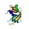
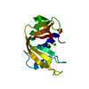
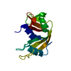
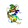
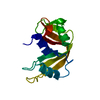
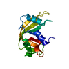
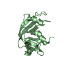
| Title | The X-ray structure of the adduct formed in the reaction between bovine pancreatic ribonuclease and carboplatin | ||||||
 Components Components | Ribonuclease pancreatic | ||||||
 Keywords Keywords | HYDROLASE/RNA / ribonuclease / RNA cleavage / HYDROLASE-RNA complex | ||||||
| Function / homology |  Function and homology information Function and homology informationpancreatic ribonuclease / ribonuclease A activity / RNA nuclease activity / nucleic acid binding / defense response to Gram-positive bacterium / hydrolase activity / extracellular region Similarity search - Function | ||||||
| Biological species |  | ||||||
| Method |  X-RAY DIFFRACTION / MOLECULAR REPLACEMENT / Resolution: 2.09 Å X-RAY DIFFRACTION / MOLECULAR REPLACEMENT / Resolution: 2.09 Å | ||||||
 Authors Authors | Merlino, A. | ||||||
 Citation Citation | Journal: J.Inorg.Biochem. / Year: 2015 Title: Interactions of carboplatin and oxaliplatin with proteins: Insights from X-ray structures and mass spectrometry studies of their ribonuclease A adducts. Authors: Messori, L. / Marzo, T. / Merlino, A. | ||||||
| History |
|
- Structure visualization
| Structure viewer | Molecule: MolmilJmol/JSmol |
|---|
- Downloads & links
-Download
| PDBx/mmCIF format | 4s0q.cif.gz | 60.9 KB | Display | PDBx/mmCIF format |
|---|---|---|---|---|
| PDB format | pdb4s0q.ent.gz | 45 KB | Display | PDB format |
| PDBx/mmJSON format | 4s0q.json.gz | Tree view | PDBx/mmJSON format | |
| Others |  Other downloads Other downloads |
-Validation report
| Arichive directory | https://data.pdbj.org/pub/pdb/validation_reports/s0/4s0qftp://data.pdbj.org/pub/pdb/validation_reports/s0/4s0q | HTTPS FTP |
|---|
-Related structure data
| Related structure data |  4s18C  1jvtS S: Starting model for refinement C: citing same article ( |
|---|---|
| Similar structure data |


-Links
 PDBj
PDBj

- Assembly
| Deposited unit | 
| ||||||||
|---|---|---|---|---|---|---|---|---|---|
| 1 | 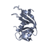
| ||||||||
| 2 | 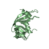
| ||||||||
| Unit cell |
|
-Components
| #1: Protein | Mass: 13708.326 Da / Num. of mol.: 2 Source method: isolated from a genetically manipulated source Source: (gene. exp.) #2: Chemical |   Mass: 371.248 Da / Num. of mol.: 2 / Source method: obtained synthetically / Formula: C6H12N2O4Pt / Comment: medication, chemotherapy*YM Mass: 371.248 Da / Num. of mol.: 2 / Source method: obtained synthetically / Formula: C6H12N2O4Pt / Comment: medication, chemotherapy*YM#3: Water | ChemComp-HOH / |  Mass: 18.015 Da / Num. of mol.: 104 / Source method: isolated from a natural source / Formula: H2O Mass: 18.015 Da / Num. of mol.: 104 / Source method: isolated from a natural source / Formula: H2OHas protein modification | Y | |
|---|
-Experimental details
-Experiment
| Experiment | Method: X-RAY DIFFRACTION / Number of used crystals: 1 |
|---|
- Sample preparation
| Crystal | Density Matthews: 2.17 Å3/Da / Density % sol: 43.21 % |
|---|---|
| Crystal grow | Temperature: 298 K / Method: vapor diffusion, hanging drop / pH: 5 Details: Crystals of the RNase A-carboplatin adduct have been obtained by soaking experiments where pre-grown monoclinic protein crystals were incubated with an excess of the platinum drug (at a ...Details: Crystals of the RNase A-carboplatin adduct have been obtained by soaking experiments where pre-grown monoclinic protein crystals were incubated with an excess of the platinum drug (at a protein to platinum ratio 1:10). Crystals of RNase A were grown by hanging-drop vapor mixing 1 L of RNase A at 20 mg mL-1 with equal volumes of reservoir solution containing 20% PEG4000 and 20 mM sodium citrate buffer pH 5.0 at 298 K. Two weeks after their appearance, crystals were soaked for four days in a solution of Carboplatin dissolved in 10 L of reservoir. At the final, Pt drug:protein concentrations were in 10:1 ratio., VAPOR DIFFUSION, HANGING DROP |
-Data collection
| Diffraction | Mean temperature: 100 K |
|---|---|
| Diffraction source | Source: ROTATING ANODE / Type: RIGAKU MICROMAX-007 HF / Wavelength: 1.5418 Å |
| Radiation | Monochromator: graphite / Protocol: SINGLE WAVELENGTH / Monochromatic (M) / Laue (L): M / Scattering type: x-ray |
| Radiation wavelength | Wavelength: 1.5418 Å / Relative weight: 1 |
| Reflection | Resolution: 2.09→72.71 Å / Num. all: 14095 / Num. obs: 14095 / % possible obs: 99.7 % / Redundancy: 5.6 % / Rmerge(I) obs: 0.124 |
- Processing
| Software | Name: REFMAC / Version: 5.8.0049 / Classification: refinement | ||||||||||||||||||||||||||||||||||||||||||||||||||||||||||||||||||||||||||||||||||||||||||||||||||||||||||||||||||||||||||||||||||||||||||||||||||||||||||||||||||||||||||||||||||||||
|---|---|---|---|---|---|---|---|---|---|---|---|---|---|---|---|---|---|---|---|---|---|---|---|---|---|---|---|---|---|---|---|---|---|---|---|---|---|---|---|---|---|---|---|---|---|---|---|---|---|---|---|---|---|---|---|---|---|---|---|---|---|---|---|---|---|---|---|---|---|---|---|---|---|---|---|---|---|---|---|---|---|---|---|---|---|---|---|---|---|---|---|---|---|---|---|---|---|---|---|---|---|---|---|---|---|---|---|---|---|---|---|---|---|---|---|---|---|---|---|---|---|---|---|---|---|---|---|---|---|---|---|---|---|---|---|---|---|---|---|---|---|---|---|---|---|---|---|---|---|---|---|---|---|---|---|---|---|---|---|---|---|---|---|---|---|---|---|---|---|---|---|---|---|---|---|---|---|---|---|---|---|---|---|
| Refinement | Method to determine structure: MOLECULAR REPLACEMENT Starting model: pdb code 1JVT, chain A without water molecules and ligands Resolution: 2.09→72.71 Å / Cor.coef. Fo:Fc: 0.945 / Cor.coef. Fo:Fc free: 0.912 / SU B: 6.663 / SU ML: 0.171 / Cross valid method: THROUGHOUT / ESU R: 0.259 / ESU R Free: 0.214 / Stereochemistry target values: MAXIMUM LIKELIHOOD / Details: HYDROGENS HAVE BEEN ADDED IN THE RIDING POSITIONS
| ||||||||||||||||||||||||||||||||||||||||||||||||||||||||||||||||||||||||||||||||||||||||||||||||||||||||||||||||||||||||||||||||||||||||||||||||||||||||||||||||||||||||||||||||||||||
| Solvent computation | Ion probe radii: 0.8 Å / Shrinkage radii: 0.8 Å / VDW probe radii: 1.2 Å / Solvent model: MASK | ||||||||||||||||||||||||||||||||||||||||||||||||||||||||||||||||||||||||||||||||||||||||||||||||||||||||||||||||||||||||||||||||||||||||||||||||||||||||||||||||||||||||||||||||||||||
| Displacement parameters | Biso mean: 37.837 Å2
| ||||||||||||||||||||||||||||||||||||||||||||||||||||||||||||||||||||||||||||||||||||||||||||||||||||||||||||||||||||||||||||||||||||||||||||||||||||||||||||||||||||||||||||||||||||||
| Refinement step | Cycle: LAST / Resolution: 2.09→72.71 Å
| ||||||||||||||||||||||||||||||||||||||||||||||||||||||||||||||||||||||||||||||||||||||||||||||||||||||||||||||||||||||||||||||||||||||||||||||||||||||||||||||||||||||||||||||||||||||
| Refine LS restraints |
|