 Movie
Movie Controller


+ Open data

- Basic information
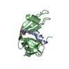
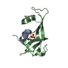
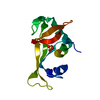
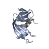
| Entry | Database: PDB / ID: 1d5h | ||||||
|---|---|---|---|---|---|---|---|
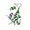
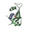
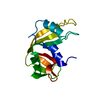
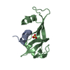
| Title | Rnase s(f8a). mutant ribonucleasE S. | ||||||
 Components Components |
| ||||||
 Keywords Keywords | HYDROLASE / RNASE S MUTANT F8A CAVITY S PROTEIN S PEPTIDE | ||||||
| Function / homology |  Function and homology information Function and homology informationpancreatic ribonuclease / ribonuclease A activity / RNA nuclease activity / nucleic acid binding / lyase activity / defense response to Gram-positive bacterium / hydrolase activity / extracellular region Similarity search - Function | ||||||
| Biological species |  | ||||||
| Method |  X-RAY DIFFRACTION / Resolution: 2.25 Å X-RAY DIFFRACTION / Resolution: 2.25 Å | ||||||
 Authors Authors | Ratnaparkhi, G.S. / Varadarajan, R. | ||||||
 Citation Citation | Journal: Biochemistry / Year: 2000 Title: Thermodynamic and structural studies of cavity formation in proteins suggest that loss of packing interactions rather than the hydrophobic effect dominates the observed energetics. Authors: Ratnaparkhi, G.S. / Varadarajan, R. #1: Journal: Proteins: Struct.,Funct.,Genet. / Year: 1999Title: X-ray crystallographic studies of the denaturation of ribonuclease S Authors: Ratnaparkhi, G.S. / Varadarajan, R. #2: Journal: Biochemistry / Year: 1992Title: Crystallographic structures of ribonuclease S variants with nonpolar substitution at position 13: packing and cavities Authors: Varadarajan, R. / Richards, F.M. #3: Journal: Biochemistry / Year: 1994Title: Thermodynamic and structural consequences of changing a sulfur atom to a methylene group in the M13Nle mutation in ribonuclease-S Authors: Thomson, J. / Ratnaparkhi, G.S. / Varadarajan, R. / Sturtevant, J.M. / Richards, F.M. | ||||||
| History |
|
- Structure visualization
| Structure viewer | Molecule: MolmilJmol/JSmol |
|---|
- Downloads & links
-Download
| PDBx/mmCIF format | 1d5h.cif.gz | 43.8 KB | Display | PDBx/mmCIF format |
|---|---|---|---|---|
| PDB format | pdb1d5h.ent.gz | 30.9 KB | Display | PDB format |
| PDBx/mmJSON format | 1d5h.json.gz | Tree view | PDBx/mmJSON format | |
| Others |  Other downloads Other downloads |
-Validation report
| Arichive directory | https://data.pdbj.org/pub/pdb/validation_reports/d5/1d5hftp://data.pdbj.org/pub/pdb/validation_reports/d5/1d5h | HTTPS FTP |
|---|
-Related structure data


-Links
 PDBj
PDBj

- Assembly
| Deposited unit | 
| ||||||||
|---|---|---|---|---|---|---|---|---|---|
| 1 |
| ||||||||
| Unit cell |
| ||||||||
| Details | Monomer. Two fragments S peptide and S protein held together by non-covalent interactions |
-Components
| #1: Protein/peptide | Mass: 1675.863 Da / Num. of mol.: 1 / Fragment: RESIDUES 1-15 / Mutation: F8A / Source method: obtained synthetically Details: S15 peptide synthesized by solid phase peptide synthesis References: UniProt: P61823 |
|---|---|
| #2: Protein | Mass: 11294.749 Da / Num. of mol.: 1 / Fragment: RESIDUES 24-124 / Source method: isolated from a natural source Details: DERIEVED FROM A LIMIT DIGEST OF BOVINE PANCREATIC RNASE A Source: (natural) |
| #3: Chemical | ChemComp-SO4 /   Mass: 96.063 Da / Num. of mol.: 1 / Source method: obtained synthetically / Formula: SO4 Mass: 96.063 Da / Num. of mol.: 1 / Source method: obtained synthetically / Formula: SO4 |
| #4: Water | ChemComp-HOH /  Mass: 18.015 Da / Num. of mol.: 35 / Source method: isolated from a natural source / Formula: H2O Mass: 18.015 Da / Num. of mol.: 35 / Source method: isolated from a natural source / Formula: H2O |
| Has protein modification | Y |
-Experimental details
-Experiment
| Experiment | Method: X-RAY DIFFRACTION / Number of used crystals: 1 |
|---|
- Sample preparation
| Crystal | Density Matthews: 2.17 Å3/Da / Density % sol: 43.28 % | ||||||||||||||||||||||||||||||
|---|---|---|---|---|---|---|---|---|---|---|---|---|---|---|---|---|---|---|---|---|---|---|---|---|---|---|---|---|---|---|---|
| Crystal grow | Temperature: 293 K / Method: vapor diffusion, sitting drop / pH: 5.75 Details: 3M CsCl, 35% Ammonium Sulfate. 0.1 M Sodium Acetate, pH 5.75, VAPOR DIFFUSION, SITTING DROP, temperature 20K | ||||||||||||||||||||||||||||||
| Crystal grow | *PLUS Method: unknown / Details: Thomson, J., (1994) Biochemistry, 33, 8587. | ||||||||||||||||||||||||||||||
| Components of the solutions | *PLUS
|
-Data collection
| Diffraction | Mean temperature: 293 K |
|---|---|
| Diffraction source | Source: ROTATING ANODE / Type: RIGAKU / Wavelength: 1.5418 |
| Detector | Type: XENTRONICS / Detector: AREA DETECTOR / Date: Mar 8, 1994 |
| Radiation | Protocol: SINGLE WAVELENGTH / Monochromatic (M) / Laue (L): M / Scattering type: x-ray |
| Radiation wavelength | Wavelength: 1.5418 Å / Relative weight: 1 |
| Reflection | Resolution: 2.2→10 Å / Num. obs: 5328 / % possible obs: 95 % / Observed criterion σ(F): 1 / Observed criterion σ(I): 1 / Redundancy: 2.5 % / Biso Wilson estimate: 22.3 Å2 / Rmerge(I) obs: 0.06 / Net I/σ(I): 13 |
| Reflection shell | Resolution: 2.25→2.37 Å / Rmerge(I) obs: 0.3 / Num. unique all: 504 / % possible all: 70 |
| Reflection | *PLUS Rmerge(I) obs: 0.06 |
- Processing
| Software |
| ||||||||||||||||||||||||||||||||||||||||||||
|---|---|---|---|---|---|---|---|---|---|---|---|---|---|---|---|---|---|---|---|---|---|---|---|---|---|---|---|---|---|---|---|---|---|---|---|---|---|---|---|---|---|---|---|---|---|
| Refinement | Resolution: 2.25→10 Å / Rfactor Rfree error: 0.012 / Data cutoff high absF: 10000000 / Data cutoff low absF: 0 / Isotropic thermal model: RESTRAINED / Cross valid method: THROUGHOUT / σ(F): 1 / Stereochemistry target values: NA / Details: NA
| ||||||||||||||||||||||||||||||||||||||||||||
| Displacement parameters | Biso mean: 13.9 Å2
| ||||||||||||||||||||||||||||||||||||||||||||
| Refine analyze |
| ||||||||||||||||||||||||||||||||||||||||||||
| Refinement step | Cycle: LAST / Resolution: 2.25→10 Å
| ||||||||||||||||||||||||||||||||||||||||||||
| Refine LS restraints |
| ||||||||||||||||||||||||||||||||||||||||||||
| LS refinement shell | Resolution: 2.25→2.37 Å / Rfactor Rfree error: 0.049 / Total num. of bins used: 7
| ||||||||||||||||||||||||||||||||||||||||||||
| Xplor file |
| ||||||||||||||||||||||||||||||||||||||||||||
| Software | *PLUS Name: X-PLOR / Version: 3.851 / Classification: refinement | ||||||||||||||||||||||||||||||||||||||||||||
| Refinement | *PLUS σ(F): 1 / % reflection Rfree: 10.6 % / Rfactor obs: 0.21 / Rfactor Rwork: 0.21 | ||||||||||||||||||||||||||||||||||||||||||||
| Solvent computation | *PLUS | ||||||||||||||||||||||||||||||||||||||||||||
| Displacement parameters | *PLUS Biso mean: 13.9 Å2 | ||||||||||||||||||||||||||||||||||||||||||||
| Refine LS restraints | *PLUS
| ||||||||||||||||||||||||||||||||||||||||||||
| LS refinement shell | *PLUS Rfactor Rfree: 0.385 / % reflection Rfree: 11 % / Rfactor Rwork: 0.305 |
