 Movie
Movie Controller

[English] 日本語
 Yorodumi
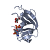
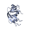
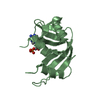
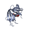
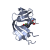
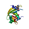
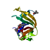
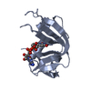
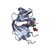
- PDB-1qhc: CRYSTAL STRUCTURE OF RIBONUCLEASE A IN COMPLEX WITH 5'-PHOSPHO-2'... -
+ Open data

- Basic information
| Entry | Database: PDB / ID: 1qhc | ||||||
|---|---|---|---|---|---|---|---|
| Title | CRYSTAL STRUCTURE OF RIBONUCLEASE A IN COMPLEX WITH 5'-PHOSPHO-2'-DEOXYURIDINE-3'-PYROPHOSPHATE ADENOSINE-3'-PHOSPHATE | ||||||
 Components Components | PROTEIN (RIBONUCLEASE A) | ||||||
 Keywords Keywords | HYDROLASE / RIBONUCLEASE / ENDONUCLEASE / NUCLEOTIDE INHIBITOR | ||||||
| Function / homology |  Function and homology information Function and homology informationpancreatic ribonuclease / ribonuclease A activity / RNA nuclease activity / nucleic acid binding / defense response to Gram-positive bacterium / hydrolase activity / extracellular region Similarity search - Function | ||||||
| Biological species |  | ||||||
| Method |  X-RAY DIFFRACTION / SYNCHROTRON / MOLECULAR REPLACEMENT / Resolution: 1.7 Å X-RAY DIFFRACTION / SYNCHROTRON / MOLECULAR REPLACEMENT / Resolution: 1.7 Å | ||||||
 Authors Authors | Leonidas, D.D. / Acharya, K.R. | ||||||
 Citation Citation | Journal: Biochemistry / Year: 1999 Title: Toward rational design of ribonuclease inhibitors: high-resolution crystal structure of a ribonuclease A complex with a potent 3',5'-pyrophosphate-linked dinucleotide inhibitor. Authors: Leonidas, D.D. / Shapiro, R. / Irons, L.I. / Russo, N. / Acharya, K.R. #1: Journal: Biochemistry / Year: 1997Title: Crystal Structures of Ribonuclease A Complexes with 5'-Diphosphoadenosine 3'- Phosphate and 5'-Diphosphoadenosine 2'-Phosphate at 1.7 A Resolution Authors: Leonidas, D.D. / Shapiro, R. / Irons, L.I. / Russo, N. / Acharya, K.R. | ||||||
| History |
|
- Structure visualization
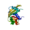
| Structure viewer | Molecule: MolmilJmol/JSmol |
|---|
- Downloads & links
-Download
| PDBx/mmCIF format | 1qhc.cif.gz | 65.9 KB | Display | PDBx/mmCIF format |
|---|---|---|---|---|
| PDB format | pdb1qhc.ent.gz | 48.5 KB | Display | PDB format |
| PDBx/mmJSON format | 1qhc.json.gz | Tree view | PDBx/mmJSON format | |
| Others |  Other downloads Other downloads |
-Validation report
| Arichive directory | https://data.pdbj.org/pub/pdb/validation_reports/qh/1qhcftp://data.pdbj.org/pub/pdb/validation_reports/qh/1qhc | HTTPS FTP |
|---|
-Related structure data
| Related structure data | 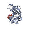 1afkS S: Starting model for refinement |
|---|---|
| Similar structure data |


-Links
 PDBj
PDBj

- Assembly
| Deposited unit | 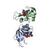
| ||||||||||
|---|---|---|---|---|---|---|---|---|---|---|---|
| 1 | 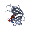
| ||||||||||
| 2 | 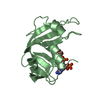
| ||||||||||
| Unit cell |
| ||||||||||
| Noncrystallographic symmetry (NCS) | NCS oper: (Code: given Matrix: (-0.574776, 0.784184, 0.233854), Vector: |
-Components
| #1: Protein | Mass: 13708.326 Da / Num. of mol.: 2 / Source method: isolated from a natural source / Source: (natural) #2: Chemical | 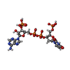  Mass: 797.348 Da / Num. of mol.: 2 / Source method: obtained synthetically / Formula: C19H27N7O20P4 Mass: 797.348 Da / Num. of mol.: 2 / Source method: obtained synthetically / Formula: C19H27N7O20P4#3: Water | ChemComp-HOH / |  Mass: 18.015 Da / Num. of mol.: 137 / Source method: isolated from a natural source / Formula: H2O Mass: 18.015 Da / Num. of mol.: 137 / Source method: isolated from a natural source / Formula: H2OHas protein modification | Y | Sequence details | 1AFK A SWS P00656 1 - 26 NOT IN ATOMS LIST 1AFK B SWS P00656 1 - 26 NOT IN ATOMS LIST | |
|---|
-Experimental details
-Experiment
| Experiment | Method: X-RAY DIFFRACTION / Number of used crystals: 1 |
|---|
- Sample preparation
| Crystal | Density Matthews: 2.2 Å3/Da / Density % sol: 48 % | ||||||||||||||||||||||||||||||
|---|---|---|---|---|---|---|---|---|---|---|---|---|---|---|---|---|---|---|---|---|---|---|---|---|---|---|---|---|---|---|---|
| Crystal grow | Method: vapor diffusion, hanging drop / pH: 5.5 Details: CRYSTALLIZED FROM 20% PEG 4000, 20 MM SODIUM CITRATE, PH 5.5, VAPOR DIFFUSION, HANGING DROP | ||||||||||||||||||||||||||||||
| Crystal grow | *PLUS Temperature: 16 ℃ / Details: Leonidas, D.D., (1997) Biochemistry, 36, 5578. | ||||||||||||||||||||||||||||||
| Components of the solutions | *PLUS
|
-Data collection
| Diffraction | Mean temperature: 293 K |
|---|---|
| Diffraction source | Source: SYNCHROTRON / Site: SRS  / Beamline: PX7.2 / Wavelength: 1.488 / Beamline: PX7.2 / Wavelength: 1.488 |
| Detector | Type: MARRESEARCH / Detector: IMAGE PLATE / Date: Jul 15, 1998 |
| Radiation | Protocol: SINGLE WAVELENGTH / Monochromatic (M) / Laue (L): M / Scattering type: x-ray |
| Radiation wavelength | Wavelength: 1.488 Å / Relative weight: 1 |
| Reflection | Resolution: 1.7→40 Å / Num. obs: 25723 / % possible obs: 94.5 % / Observed criterion σ(I): 0 / Redundancy: 3 % / Biso Wilson estimate: 25.8 Å2 / Rsym value: 0.083 / Net I/σ(I): 5.7 |
| Reflection shell | Resolution: 1.7→1.76 Å / Redundancy: 1.8 % / Mean I/σ(I) obs: 2.05 / Rsym value: 0.326 / % possible all: 84 |
- Processing
| Software |
| ||||||||||||||||||||||||||||||||||||||||||||||||||||||||||||||||||||||||||||||||
|---|---|---|---|---|---|---|---|---|---|---|---|---|---|---|---|---|---|---|---|---|---|---|---|---|---|---|---|---|---|---|---|---|---|---|---|---|---|---|---|---|---|---|---|---|---|---|---|---|---|---|---|---|---|---|---|---|---|---|---|---|---|---|---|---|---|---|---|---|---|---|---|---|---|---|---|---|---|---|---|---|---|
| Refinement | Method to determine structure: MOLECULAR REPLACEMENT Starting model: PDB ENTRY 1AFK Resolution: 1.7→20 Å / Rfactor Rfree error: 0.007 / Data cutoff high absF: 10000000 / Data cutoff low absF: 0.001 / Isotropic thermal model: RESTRAINED / Cross valid method: THROUGHOUT / σ(F): 0 Details: BULK SOLVENT MODEL USED DISORDERED REGIONS WERE MODELED STEREOCHEMICALLY.
| ||||||||||||||||||||||||||||||||||||||||||||||||||||||||||||||||||||||||||||||||
| Displacement parameters | Biso mean: 32.1 Å2 | ||||||||||||||||||||||||||||||||||||||||||||||||||||||||||||||||||||||||||||||||
| Refine analyze |
| ||||||||||||||||||||||||||||||||||||||||||||||||||||||||||||||||||||||||||||||||
| Refinement step | Cycle: LAST / Resolution: 1.7→20 Å
| ||||||||||||||||||||||||||||||||||||||||||||||||||||||||||||||||||||||||||||||||
| Refine LS restraints |
| ||||||||||||||||||||||||||||||||||||||||||||||||||||||||||||||||||||||||||||||||
| LS refinement shell | Resolution: 1.7→1.81 Å / Rfactor Rfree error: 0.029 / Total num. of bins used: 6
| ||||||||||||||||||||||||||||||||||||||||||||||||||||||||||||||||||||||||||||||||
| Xplor file | Serial no: 1 / Param file: PROTEIN_REP.PARAM / Topol file: TOPHCSDX.PRO | ||||||||||||||||||||||||||||||||||||||||||||||||||||||||||||||||||||||||||||||||
| Software | *PLUS Name: X-PLOR / Version: 3.851 / Classification: refinement | ||||||||||||||||||||||||||||||||||||||||||||||||||||||||||||||||||||||||||||||||
| Refinement | *PLUS σ(F): 0 / % reflection Rfree: 5 % / Rfactor Rfree: 0.26 / Rfactor Rwork: 0.2 | ||||||||||||||||||||||||||||||||||||||||||||||||||||||||||||||||||||||||||||||||
| Solvent computation | *PLUS | ||||||||||||||||||||||||||||||||||||||||||||||||||||||||||||||||||||||||||||||||
| Displacement parameters | *PLUS Biso mean: 32.1 Å2 | ||||||||||||||||||||||||||||||||||||||||||||||||||||||||||||||||||||||||||||||||
| Refine LS restraints | *PLUS
| ||||||||||||||||||||||||||||||||||||||||||||||||||||||||||||||||||||||||||||||||
| LS refinement shell | *PLUS Rfactor Rfree: 0.386 / % reflection Rfree: 4.9 % / Rfactor Rwork: 0.374 |
