 Movie
Movie Controller

[English] 日本語
 Yorodumi
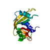
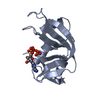
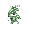
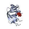
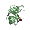
- PDB-1rob: STRUCTURE OF THE CRYSTALLINE COMPLEX OF CYTIDYLIC ACID (2'-CMP) W... -
+ Open data

- Basic information
| Entry | Database: PDB / ID: 1rob | ||||||
|---|---|---|---|---|---|---|---|
| Title | STRUCTURE OF THE CRYSTALLINE COMPLEX OF CYTIDYLIC ACID (2'-CMP) WITH RIBONUCLEASE AT 1.6 ANGSTROMS RESOLUTION | ||||||
 Components Components | RIBONUCLEASE A | ||||||
 Keywords Keywords | HYDROLASE(ENDORIBONUCLEASE) | ||||||
| Function / homology |  Function and homology information Function and homology informationpancreatic ribonuclease / ribonuclease A activity / RNA nuclease activity / nucleic acid binding / defense response to Gram-positive bacterium / hydrolase activity / extracellular region Similarity search - Function | ||||||
| Biological species |  | ||||||
| Method |  X-RAY DIFFRACTION / Resolution: 1.6 Å X-RAY DIFFRACTION / Resolution: 1.6 Å | ||||||
 Authors Authors | Lisgarten, J.N. / Palmer, R.A. | ||||||
 Citation Citation | Journal: Acta Crystallogr.,Sect.D / Year: 1993 Title: Structure of the crystalline complex of cytidylic acid (2'-CMP) with ribonuclease at 1.6 A resolution. Conservation of solvent sites in RNase-A high-resolution structures. Authors: Lisgarten, J.N. / Gupta, V. / Maes, D. / Wyns, L. / Zegers, I. / Palmer, R.A. / Dealwis, C.G. / Aguilar, C.F. / Hemmings, A.M. #1: Journal: J.Mol.Biol. / Year: 1991Title: Newly Observed Binding Mode in Pancreatic Ribonuclease Authors: Aguilar, C.F. / Thomas, P.J. / Mills, A. / Moss, D.S. / Palmer, R.A. #2: Journal: J.Mol.Biol. / Year: 1987Title: X-Ray Refinement Study on the Binding of Cytidylic Acid (2'-Cmp) to Ribonuclease A Authors: Howlin, B. / Harris, W. / Moss, D.S. / Palmer, R.A. #3: Journal: J.Crystallogr.Spectrosc.Res. / Year: 1984Title: The Refined Structure of Ribonuclease-A at 1.45 Angstroms Resolution Authors: Borkakoti, N. / Moss, D.S. / Stanford, M.J. / Palmer, R.A. | ||||||
| History |
|
- Structure visualization
| Structure viewer | Molecule: MolmilJmol/JSmol |
|---|
- Downloads & links
-Download
| PDBx/mmCIF format | 1rob.cif.gz | 37.7 KB | Display | PDBx/mmCIF format |
|---|---|---|---|---|
| PDB format | pdb1rob.ent.gz | 25.2 KB | Display | PDB format |
| PDBx/mmJSON format | 1rob.json.gz | Tree view | PDBx/mmJSON format | |
| Others |  Other downloads Other downloads |
-Validation report
| Arichive directory | https://data.pdbj.org/pub/pdb/validation_reports/ro/1robftp://data.pdbj.org/pub/pdb/validation_reports/ro/1rob | HTTPS FTP |
|---|
-Related structure data
| Similar structure data |
|---|


-Links
 PDBj
PDBj

- Assembly
| Deposited unit | 
| ||||||||
|---|---|---|---|---|---|---|---|---|---|
| 1 |
| ||||||||
| Unit cell |
| ||||||||
| Atom site foot note | 1: CIS PROLINE - PRO 93 / 2: CIS PROLINE - PRO 114 |
-Components
| #1: Protein | Mass: 13708.326 Da / Num. of mol.: 1 Source method: isolated from a genetically manipulated source Source: (gene. exp.) |
|---|---|
| #2: Chemical | ChemComp-C2P /   Mass: 323.197 Da / Num. of mol.: 1 / Source method: obtained synthetically / Formula: C9H14N3O8P Mass: 323.197 Da / Num. of mol.: 1 / Source method: obtained synthetically / Formula: C9H14N3O8P |
| #3: Water | ChemComp-HOH /  Mass: 18.015 Da / Num. of mol.: 101 / Source method: isolated from a natural source / Formula: H2O Mass: 18.015 Da / Num. of mol.: 101 / Source method: isolated from a natural source / Formula: H2O |
| Has protein modification | Y |
-Experimental details
-Experiment
| Experiment | Method: X-RAY DIFFRACTION |
|---|
- Sample preparation
| Crystal | Density Matthews: 2.18 Å3/Da / Density % sol: 43.55 % |
|---|---|
| Crystal grow | *PLUS Method: unknown / PH range low: 5.7 / PH range high: 5.2 |
| Components of the solutions | *PLUS Conc.: 30-40 % / Common name: ethanol |
-Data collection
| Radiation | Scattering type: x-ray |
|---|---|
| Radiation wavelength | Relative weight: 1 |
| Reflection | *PLUS Num. obs: 11945 / Num. measured all: 39576 |
| Reflection shell | *PLUS Highest resolution: 1.54 Å / Lowest resolution: 1.63 Å / % possible obs: 76 % / Rmerge(I) obs: 0.227 |
- Processing
| Software | Name: RESTRAIN / Classification: refinement | ||||||||||||
|---|---|---|---|---|---|---|---|---|---|---|---|---|---|
| Refinement | Rfactor obs: 0.17 / Highest resolution: 1.6 Å | ||||||||||||
| Refinement step | Cycle: LAST / Highest resolution: 1.6 Å
| ||||||||||||
| Refine LS restraints |
| ||||||||||||
| Software | *PLUS Name: RESTRAIN / Classification: refinement | ||||||||||||
| Refinement | *PLUS Highest resolution: 1.6 Å / Lowest resolution: 20 Å / Num. reflection obs: 11945 / Rfactor obs: 0.17 | ||||||||||||
| Solvent computation | *PLUS | ||||||||||||
| Displacement parameters | *PLUS |
