 Movie
Movie Controller

[English] 日本語
 Yorodumi
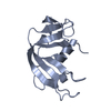
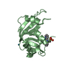
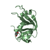
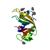
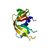
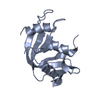
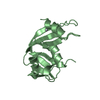
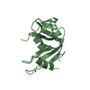
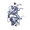
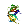
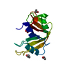
- PDB-1eic: CRYSTAL STRUCTURE OF F120A MUTANT OF BOVINE PANCREATIC RIBONUCLEASE A -
+ Open data

- Basic information
| Entry | Database: PDB / ID: 1eic | ||||||
|---|---|---|---|---|---|---|---|
| Title | CRYSTAL STRUCTURE OF F120A MUTANT OF BOVINE PANCREATIC RIBONUCLEASE A | ||||||
 Components Components | RIBONUCLEASE A | ||||||
 Keywords Keywords | HYDROLASE / ribonuclease / RNase A / Bovine pancreas | ||||||
| Function / homology |  Function and homology information Function and homology informationpancreatic ribonuclease / ribonuclease A activity / RNA nuclease activity / nucleic acid binding / defense response to Gram-positive bacterium / hydrolase activity / extracellular region Similarity search - Function | ||||||
| Biological species |  | ||||||
| Method |  X-RAY DIFFRACTION / SYNCHROTRON / Resolution: 1.4 Å X-RAY DIFFRACTION / SYNCHROTRON / Resolution: 1.4 Å | ||||||
 Authors Authors | Chatani, E. / Hayashi, R. / Moriyama, H. / Ueki, T. | ||||||
 Citation Citation | Journal: Protein Sci. / Year: 2002 Title: Conformational strictness required for maximum activity and stability of bovine pancreatic ribonuclease A as revealed by crystallographic study of three Phe120 mutants at 1.4 A resolution. Authors: Chatani, E. / Hayashi, R. / Moriyama, H. / Ueki, T. | ||||||
| History |
|
- Structure visualization
| Structure viewer | Molecule: MolmilJmol/JSmol |
|---|
- Downloads & links
-Download
| PDBx/mmCIF format | 1eic.cif.gz | 41.1 KB | Display | PDBx/mmCIF format |
|---|---|---|---|---|
| PDB format | pdb1eic.ent.gz | 28.2 KB | Display | PDB format |
| PDBx/mmJSON format | 1eic.json.gz | Tree view | PDBx/mmJSON format | |
| Others |  Other downloads Other downloads |
-Validation report
| Arichive directory | https://data.pdbj.org/pub/pdb/validation_reports/ei/1eicftp://data.pdbj.org/pub/pdb/validation_reports/ei/1eic | HTTPS FTP |
|---|
-Related structure data
| Related structure data | 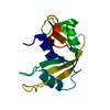 1eidC 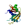 1eieC 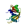 1fs3C  1dp1 C: citing same article ( |
|---|---|
| Similar structure data |


-Links
 PDBj
PDBj

- Assembly
| Deposited unit | 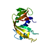
| ||||||||
|---|---|---|---|---|---|---|---|---|---|
| 1 |
| ||||||||
| Unit cell |
| ||||||||
| Details | The biological assembly is a monomer. |
-Components
| #1: Protein | Mass: 13632.229 Da / Num. of mol.: 1 / Mutation: F120A Source method: isolated from a genetically manipulated source Source: (gene. exp.)  |
|---|---|
| #2: Water | ChemComp-HOH /  Mass: 18.015 Da / Num. of mol.: 220 / Source method: isolated from a natural source / Formula: H2O Mass: 18.015 Da / Num. of mol.: 220 / Source method: isolated from a natural source / Formula: H2O |
| Has protein modification | Y |
-Experimental details
-Experiment
| Experiment | Method: X-RAY DIFFRACTION / Number of used crystals: 1 |
|---|
- Sample preparation
| Crystal | Density Matthews: 2.72 Å3/Da / Density % sol: 54.84 % | |||||||||||||||||||||||||||||||||||
|---|---|---|---|---|---|---|---|---|---|---|---|---|---|---|---|---|---|---|---|---|---|---|---|---|---|---|---|---|---|---|---|---|---|---|---|---|
| Crystal grow | Temperature: 298 K / Method: vapor diffusion, hanging drop / pH: 6 Details: ammonium sulfate, sodium chloride, sodium acetate, pH 6.0, VAPOR DIFFUSION, HANGING DROP, temperature 298K | |||||||||||||||||||||||||||||||||||
| Crystal grow | *PLUS Temperature: 25 ℃ | |||||||||||||||||||||||||||||||||||
| Components of the solutions | *PLUS
|
-Data collection
| Diffraction | Mean temperature: 100 K |
|---|---|
| Diffraction source | Source: SYNCHROTRON / Site: SPring-8  / Beamline: BL44B2 / Wavelength: 0.7 / Beamline: BL44B2 / Wavelength: 0.7 |
| Detector | Type: ADSC QUANTUM 4r / Detector: CCD / Date: Jun 3, 1999 |
| Radiation | Protocol: SINGLE WAVELENGTH / Monochromatic (M) / Laue (L): M / Scattering type: x-ray |
| Radiation wavelength | Wavelength: 0.7 Å / Relative weight: 1 |
| Reflection | Resolution: 1→16.8 Å / Num. all: 80127 / Num. obs: 80089 / % possible obs: 99.7 % / Observed criterion σ(I): 2 / Biso Wilson estimate: 11.5 Å2 / Rmerge(I) obs: 0.07 / Net I/σ(I): 36.94 |
| Reflection shell | Resolution: 1→1.008 Å / Num. unique all: 1923 |
| Reflection | *PLUS Num. measured all: 1446353 |
- Processing
| Software |
| ||||||||||||||||||||||||||||||||||||||||||||
|---|---|---|---|---|---|---|---|---|---|---|---|---|---|---|---|---|---|---|---|---|---|---|---|---|---|---|---|---|---|---|---|---|---|---|---|---|---|---|---|---|---|---|---|---|---|
| Refinement | Resolution: 1.4→5 Å / Rfactor Rfree error: 0.005 / Data cutoff high absF: 108876.72 / Data cutoff low absF: 0 / Isotropic thermal model: RESTRAINED / Cross valid method: THROUGHOUT / σ(F): 2
| ||||||||||||||||||||||||||||||||||||||||||||
| Displacement parameters | Biso mean: 17.6 Å2
| ||||||||||||||||||||||||||||||||||||||||||||
| Refine analyze |
| ||||||||||||||||||||||||||||||||||||||||||||
| Refinement step | Cycle: LAST / Resolution: 1.4→5 Å
| ||||||||||||||||||||||||||||||||||||||||||||
| Refine LS restraints |
| ||||||||||||||||||||||||||||||||||||||||||||
| LS refinement shell | Resolution: 1.4→1.45 Å / Rfactor Rfree error: 0.014 / Total num. of bins used: 10
| ||||||||||||||||||||||||||||||||||||||||||||
| Xplor file |
| ||||||||||||||||||||||||||||||||||||||||||||
| Software | *PLUS Name: X-PLOR / Version: 3.851 / Classification: refinement | ||||||||||||||||||||||||||||||||||||||||||||
| Refinement | *PLUS σ(F): 2 / % reflection Rfree: 10.3 % / Rfactor obs: 0.201 | ||||||||||||||||||||||||||||||||||||||||||||
| Solvent computation | *PLUS | ||||||||||||||||||||||||||||||||||||||||||||
| Displacement parameters | *PLUS Biso mean: 17.6 Å2 | ||||||||||||||||||||||||||||||||||||||||||||
| Refine LS restraints | *PLUS
| ||||||||||||||||||||||||||||||||||||||||||||
| LS refinement shell | *PLUS Rfactor Rfree: 0.243 / % reflection Rfree: 9.9 % / Rfactor Rwork: 0.242 |
