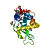
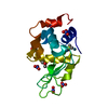
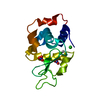
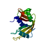
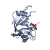
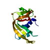
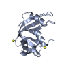
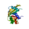
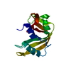
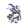
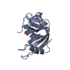
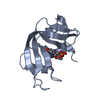
- PDB-6gok: X-ray structure of the adduct formed upon reaction of bovine panc... -
+
Open data
ID or keywords:
Loading...
-
Basic information
Entry
Database: PDB / ID: 6gok
Title
X-ray structure of the adduct formed upon reaction of bovine pancreatic ribonuclease with a Pd(II) complex bearing N,N-pyridylbenzimidazole derivative with an alkylated sulphonate side chain
Components
Ribonuclease pancreatic
Keywords
HYDROLASE / protein-metallodrug interaction / palladium-based drug / N-pyridylbenzimidazole bidentate ligands / stacking / non covalent bond
Function / homology
Function and homology information
pancreatic ribonuclease / ribonuclease A activity / RNA nuclease activity / nucleic acid binding / defense response to Gram-positive bacterium / hydrolase activity / extracellular region Similarity search - Function
P-30 Protein / Ribonuclease A-like domain / Pancreatic ribonuclease / Ribonuclease A, active site / Ribonuclease A-domain / Ribonuclease A-like domain superfamily / Pancreatic ribonuclease / Pancreatic ribonuclease family signature. / Pancreatic ribonuclease / Roll / Alpha Beta Similarity search - Domain/homology
Resolution: 2.65→72.66 Å / Cor.coef. Fo:Fc: 0.944 / Cor.coef. Fo:Fc free: 0.889 / SU B: 14.123 / SU ML: 0.286 / Cross valid method: THROUGHOUT / ESU R Free: 0.404 / Stereochemistry target values: MAXIMUM LIKELIHOOD / Details: HYDROGENS HAVE BEEN ADDED IN THE RIDING POSITIONS
Rfactor
Num. reflection
% reflection
Selection details
Rfree
0.25476
309
4.8 %
RANDOM
Rwork
0.18722
-
-
-
obs
0.19066
6196
91.22 %
-
Solvent computation
Ion probe radii: 0.8 Å / Shrinkage radii: 0.8 Å / VDW probe radii: 1.2 Å / Solvent model: MASK
 Movie
Movie Controller

 Yorodumi
 Open data
 Basic information
 Components
 Keywords
 Function and homology information
 X-RAY DIFFRACTION /
  Authors
 Citation
 Structure visualization
 Downloads & links
 Other downloads
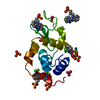


 PDBj
PDBj

 Assembly
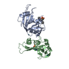
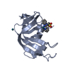
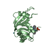

 Mass: 106.420 Da / Num. of mol.: 2 / Source method: obtained synthetically / Formula: Pd
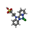
 Mass: 494.689 Da / Num. of mol.: 2 / Source method: obtained synthetically / Formula: C15H15Cl2N3O3PdS
 Mass: 18.015 Da / Num. of mol.: 63 / Source method: isolated from a natural source / Formula: H2O
 Sample preparation
 Processing