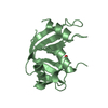
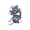
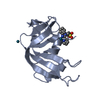
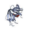
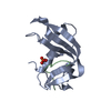
Entry Database : PDB / ID : 5oldTitle X-ray structure of the adduct formed upon reaction of ribonuclease A with a tetranuclear Pt-thiosemicarbazone compound Ribonuclease pancreatic Keywords / / / / Function / homology Function Domain/homology Component
/ / / / / / / / / / / / / / / / Biological species Bos taurus (domestic cattle)Method / / Resolution : 1.78 Å Authors Ferraro, G. / Merlino, A. Journal : J. Inorg. Biochem. / Year : 2018Title : Reactions of a tetranuclear Pt-thiosemicarbazone complex with model proteins.Authors : Marzo, T. / Navas, F. / Cirri, D. / Merlino, A. / Ferraro, G. / Messori, L. / Quiroga, A.G. History Deposition Jul 27, 2017 Deposition site / Processing site Revision 1.0 Aug 29, 2018 Provider / Type Revision 1.1 Mar 13, 2019 Group / Database references / Category / citation_author / pdbx_database_procItem _citation.country / _citation.journal_abbrev ... _citation.country / _citation.journal_abbrev / _citation.journal_id_ASTM / _citation.journal_id_CSD / _citation.journal_id_ISSN / _citation.journal_volume / _citation.page_first / _citation.page_last / _citation.pdbx_database_id_DOI / _citation.pdbx_database_id_PubMed / _citation.title / _citation.year / _citation_author.name Revision 1.2 Jan 17, 2024 Group Data collection / Database references ... Data collection / Database references / Derived calculations / Refinement description Category chem_comp_atom / chem_comp_bond ... chem_comp_atom / chem_comp_bond / database_2 / pdbx_initial_refinement_model / pdbx_struct_conn_angle / struct_conn Item _database_2.pdbx_DOI / _database_2.pdbx_database_accession ... _database_2.pdbx_DOI / _database_2.pdbx_database_accession / _pdbx_struct_conn_angle.ptnr1_auth_comp_id / _pdbx_struct_conn_angle.ptnr1_auth_seq_id / _pdbx_struct_conn_angle.ptnr1_label_asym_id / _pdbx_struct_conn_angle.ptnr1_label_atom_id / _pdbx_struct_conn_angle.ptnr1_label_comp_id / _pdbx_struct_conn_angle.ptnr3_auth_comp_id / _pdbx_struct_conn_angle.ptnr3_auth_seq_id / _pdbx_struct_conn_angle.ptnr3_label_asym_id / _pdbx_struct_conn_angle.ptnr3_label_atom_id / _pdbx_struct_conn_angle.ptnr3_label_comp_id / _pdbx_struct_conn_angle.value / _struct_conn.pdbx_dist_value / _struct_conn.pdbx_ptnr1_label_alt_id / _struct_conn.ptnr1_auth_asym_id / _struct_conn.ptnr1_auth_comp_id / _struct_conn.ptnr1_auth_seq_id / _struct_conn.ptnr1_label_asym_id / _struct_conn.ptnr1_label_atom_id / _struct_conn.ptnr1_label_comp_id / _struct_conn.ptnr1_label_seq_id / _struct_conn.ptnr2_auth_asym_id / _struct_conn.ptnr2_auth_comp_id / _struct_conn.ptnr2_auth_seq_id / _struct_conn.ptnr2_label_asym_id / _struct_conn.ptnr2_label_atom_id / _struct_conn.ptnr2_label_comp_id Revision 1.3 Nov 20, 2024 Group / Category / pdbx_modification_feature
Show all Show less
 Movie
Movie Controller

 Yorodumi
 Open data
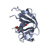
 Basic information
 Components
 Keywords
 Function and homology information
 X-RAY DIFFRACTION /
  Authors
 Citation
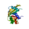
 Structure visualization
 Downloads & links
 Other downloads


 PDBj
PDBj


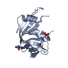
 Assembly
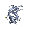
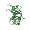

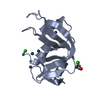
 Mass: 195.078 Da / Num. of mol.: 4 / Source method: obtained synthetically / Formula: Pt / Feature type: SUBJECT OF INVESTIGATION
 Mass: 78.133 Da / Num. of mol.: 5 / Source method: obtained synthetically / Formula: C2H6OS / Comment: DMSO, precipitant*YM
 Mass: 18.015 Da / Num. of mol.: 209 / Source method: isolated from a natural source / Formula: H2O
 Sample preparation
 Processing