 Movie
Movie Controller

[English] 日本語
 Yorodumi
- PDB-1r3m: Crystal structure of the dimeric unswapped form of bovine seminal... -
+ Open data

- Basic information
| Entry | Database: PDB / ID: 1r3m | ||||||
|---|---|---|---|---|---|---|---|
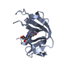
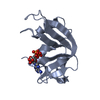
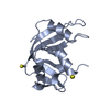
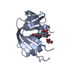
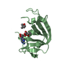
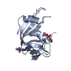
| Title | Crystal structure of the dimeric unswapped form of bovine seminal ribonuclease | ||||||
 Components Components | Ribonuclease, seminal | ||||||
 Keywords Keywords | HYDROLASE / RIBONUCLEASE / SWAPPING / HINGE | ||||||
| Function / homology |  Function and homology information Function and homology informationpancreatic ribonuclease / ribonuclease A activity / RNA nuclease activity / nucleic acid binding / defense response to Gram-positive bacterium / hydrolase activity / extracellular region / identical protein binding Similarity search - Function | ||||||
| Biological species |  | ||||||
| Method |  X-RAY DIFFRACTION / SYNCHROTRON / MOLECULAR REPLACEMENT / Resolution: 2.2 Å X-RAY DIFFRACTION / SYNCHROTRON / MOLECULAR REPLACEMENT / Resolution: 2.2 Å | ||||||
 Authors Authors | Berisio, R. / Sica, F. / De Lorenzo, C. / Di Fiore, A. / Piccoli, R. / Zagari, A. / Mazzarella, L. | ||||||
 Citation Citation | Journal: Febs Lett. / Year: 2003 Title: Crystal structure of the dimeric unswapped form of bovine seminal ribonuclease Authors: Berisio, R. / Sica, F. / De Lorenzo, C. / Di Fiore, A. / Piccoli, R. / Zagari, A. / Mazzarella, L. #1: Journal: Acta Crystallogr.,Sect.D / Year: 1993Title: BOVINE SEMINAL RIBONUCLEASE: STRUCTURE AT 1.9 A RESOLUTION Authors: Mazzarella, L. / CAPASSO, S. / DE MASI, D. / DI LORENZO, G. / MATTIA, C.A. / ZAGARI, A. #2: Journal: Proteins / Year: 2003Title: THE UNSWAPPED CHAIN OF BOVINE SEMINAL RIBONUCLEASE: CRYSTAL STRUCTURE OF THE FREE AND LIGANDED MONOMERIC DERIVATIVE Authors: SICA, F. / DI FIORE, A. / ZAGARI, A. / MAZZARELLA, L. | ||||||
| History |
|
- Structure visualization
| Structure viewer | Molecule: MolmilJmol/JSmol |
|---|
- Downloads & links
-Download
| PDBx/mmCIF format | 1r3m.cif.gz | 60 KB | Display | PDBx/mmCIF format |
|---|---|---|---|---|
| PDB format | pdb1r3m.ent.gz | 44.3 KB | Display | PDB format |
| PDBx/mmJSON format | 1r3m.json.gz | Tree view | PDBx/mmJSON format | |
| Others |  Other downloads Other downloads |
-Validation report
| Arichive directory | https://data.pdbj.org/pub/pdb/validation_reports/r3/1r3mftp://data.pdbj.org/pub/pdb/validation_reports/r3/1r3m | HTTPS FTP |
|---|
-Related structure data
| Related structure data | 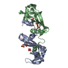 1bsrS S: Starting model for refinement |
|---|---|
| Similar structure data |


-Links
 PDBj
PDBj

- Assembly
| Deposited unit | 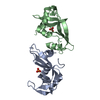
| ||||||||
|---|---|---|---|---|---|---|---|---|---|
| 1 | 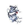
| ||||||||
| 2 | 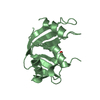
| ||||||||
| Unit cell |
|
-Components
| #1: Protein | Mass: 13632.640 Da / Num. of mol.: 2 / Source method: isolated from a natural source / Source: (natural) #2: Chemical |   Mass: 94.971 Da / Num. of mol.: 2 / Source method: obtained synthetically / Formula: PO4 Mass: 94.971 Da / Num. of mol.: 2 / Source method: obtained synthetically / Formula: PO4#3: Water | ChemComp-HOH / |  Mass: 18.015 Da / Num. of mol.: 101 / Source method: isolated from a natural source / Formula: H2O Mass: 18.015 Da / Num. of mol.: 101 / Source method: isolated from a natural source / Formula: H2OHas protein modification | Y | |
|---|
-Experimental details
-Experiment
| Experiment | Method: X-RAY DIFFRACTION |
|---|
- Sample preparation
| Crystal | Density Matthews: 2.49 Å3/Da / Density % sol: 50.59 % | ||||||||||||||||||||||||||||||
|---|---|---|---|---|---|---|---|---|---|---|---|---|---|---|---|---|---|---|---|---|---|---|---|---|---|---|---|---|---|---|---|
| Crystal grow | Temperature: 294 K / Method: evaporation / pH: 8.4 Details: MPD, ammonium sulfate, pH 8.4, EVAPORATION, temperature 294K | ||||||||||||||||||||||||||||||
| Crystal grow | *PLUS Method: vapor diffusion, sitting drop | ||||||||||||||||||||||||||||||
| Components of the solutions | *PLUS
|
-Data collection
| Diffraction | Mean temperature: 100 K |
|---|---|
| Diffraction source | Source: SYNCHROTRON / Site: ELETTRA  / Beamline: 5.2R / Wavelength: 1 Å / Beamline: 5.2R / Wavelength: 1 Å |
| Detector | Type: MARRESEARCH / Detector: IMAGE PLATE / Date: Sep 5, 1998 |
| Radiation | Monochromator: NULL / Protocol: SINGLE WAVELENGTH / Monochromatic (M) / Laue (L): M / Scattering type: x-ray |
| Radiation wavelength | Wavelength: 1 Å / Relative weight: 1 |
| Reflection | Resolution: 2.2→15 Å / Num. all: 13585 / Num. obs: 13585 / % possible obs: 99.3 % / Observed criterion σ(F): 0 / Observed criterion σ(I): 0 / Redundancy: 3.9 % / Biso Wilson estimate: 20 Å2 / Rmerge(I) obs: 0.048 / Net I/σ(I): 29.4 |
| Reflection shell | Resolution: 2.2→2.24 Å / Redundancy: 2.4 % / Rmerge(I) obs: 0.086 / Mean I/σ(I) obs: 15.1 / Num. unique all: 2000 / % possible all: 99.9 |
| Reflection | *PLUS Lowest resolution: 15 Å |
| Reflection shell | *PLUS Highest resolution: 2.2 Å / % possible obs: 99.9 % |
- Processing
| Software |
| |||||||||||||||||||||||||
|---|---|---|---|---|---|---|---|---|---|---|---|---|---|---|---|---|---|---|---|---|---|---|---|---|---|---|
| Refinement | Method to determine structure: MOLECULAR REPLACEMENT Starting model: PDB ENTRY 1BSR Resolution: 2.2→15 Å / Isotropic thermal model: Isotropic / Cross valid method: THROUGHOUT / σ(F): 2 / Stereochemistry target values: Engh & Huber
| |||||||||||||||||||||||||
| Displacement parameters | Biso mean: 31.3 Å2 | |||||||||||||||||||||||||
| Refinement step | Cycle: LAST / Resolution: 2.2→15 Å
| |||||||||||||||||||||||||
| LS refinement shell | Resolution: 2.2→2.3 Å /
| |||||||||||||||||||||||||
| Refinement | *PLUS Lowest resolution: 15 Å | |||||||||||||||||||||||||
| Solvent computation | *PLUS | |||||||||||||||||||||||||
| Displacement parameters | *PLUS |
