 Movie
Movie Controller

[English] 日本語
 Yorodumi
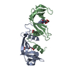
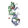
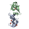
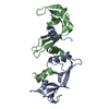
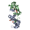
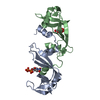
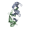
- PDB-1r5c: X-ray structure of the complex of Bovine seminal ribonuclease swa... -
+ Open data

- Basic information
| Entry | Database: PDB / ID: 1r5c | ||||||
|---|---|---|---|---|---|---|---|
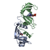
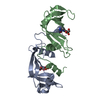
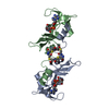
| Title | X-ray structure of the complex of Bovine seminal ribonuclease swapping dimer with d(CpA) | ||||||
 Components Components | Ribonuclease, seminal | ||||||
 Keywords Keywords | HYDROLASE / ribonucleases / protein dynamics / protein structure-function / ligand binding / population shift / 3D domain swapping | ||||||
| Function / homology |  Function and homology information Function and homology informationpancreatic ribonuclease / ribonuclease A activity / RNA nuclease activity / nucleic acid binding / defense response to Gram-positive bacterium / hydrolase activity / extracellular region / identical protein binding Similarity search - Function | ||||||
| Biological species |  | ||||||
| Method |  X-RAY DIFFRACTION / MOLECULAR REPLACEMENT / Resolution: 2.1 Å X-RAY DIFFRACTION / MOLECULAR REPLACEMENT / Resolution: 2.1 Å | ||||||
 Authors Authors | Merlino, A. / Vitagliano, L. / Sica, F. / Zagari, A. / Mazzarella, L. | ||||||
 Citation Citation | Journal: Biopolymers / Year: 2004 Title: Population shift vs induced fit: The case of bovine seminal ribonuclease swapping dimer Authors: Merlino, A. / Vitagliano, L. / Sica, F. / Zagari, A. / Mazzarella, L. #1: Journal: Acta Crystallogr.,Sect.D / Year: 1993Title: BOVINE SEMINAL RIBONUCLEASE: STRUCTURE AT 1.9 A RESOLUTION Authors: Mazzarella, L. / Capasso, S. / Demasi, D. / Di Lorenzo, G. / Mattia, C.A. / Zagari, A. #2: Journal: Protein Sci. / Year: 1998Title: Binding of a Substrate Analog to a Domain Swapping Protein: X-Ray Structure of the Complex of Bovine Seminal Ribonuclease with Uridylyl(2'-5')Adenosine Authors: Vitagliano, L. / Adinolfi, S. / Riccio, A. / Sica, F. / Zagari, A. / Mazzarella, L. #3: Journal: J.Cryst.Growth / Year: 1996Title: Cosolute Effect on Crystallization of Two Dinucleotide Complexes of Bovine Seminal Ribonuclease from Concentrated Salt Solutions Authors: Sica, F. / Adinolfi, S. / Vitagliano, L. / Zagari, A. / Capasso, S. / Mazzarella, L. #4: Journal: J.Mol.Biol. / Year: 1999Title: A potential allosteric subsite generated by domain swapping in bovine seminal ribonuclease Authors: VITAGLIANO, L. / Adinolfi, S. / Sica, F. / Merlino, A. / Zagari, A. / Mazzarella, L. #5: Journal: J.Cryst.Growth / Year: 1999Title: Crystallization of Multiple Forms of Bovine Seminal Ribonuclease: the liganded and Unliganded State | ||||||
| History |
|
- Structure visualization
| Structure viewer | Molecule: MolmilJmol/JSmol |
|---|
- Downloads & links
-Download
| PDBx/mmCIF format | 1r5c.cif.gz | 62.3 KB | Display | PDBx/mmCIF format |
|---|---|---|---|---|
| PDB format | pdb1r5c.ent.gz | 46.3 KB | Display | PDB format |
| PDBx/mmJSON format | 1r5c.json.gz | Tree view | PDBx/mmJSON format | |
| Others |  Other downloads Other downloads |
-Validation report
| Arichive directory | https://data.pdbj.org/pub/pdb/validation_reports/r5/1r5cftp://data.pdbj.org/pub/pdb/validation_reports/r5/1r5c | HTTPS FTP |
|---|
-Related structure data
| Related structure data | 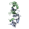 1r5dC  1bsrS C: citing same article ( S: Starting model for refinement |
|---|---|
| Similar structure data |
-Links
 PDBj
PDBj

- Assembly
| Deposited unit | 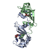
| ||||||||
|---|---|---|---|---|---|---|---|---|---|
| 1 |
| ||||||||
| Unit cell |
|
-Components
| #1: Protein | Mass: 13632.640 Da / Num. of mol.: 2 / Source method: isolated from a natural source / Source: (natural) #2: Chemical | 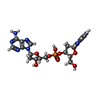  Mass: 540.424 Da / Num. of mol.: 2 / Source method: obtained synthetically / Formula: C19H25N8O9P Mass: 540.424 Da / Num. of mol.: 2 / Source method: obtained synthetically / Formula: C19H25N8O9P#3: Water | ChemComp-HOH / |  Mass: 18.015 Da / Num. of mol.: 91 / Source method: isolated from a natural source / Formula: H2O Mass: 18.015 Da / Num. of mol.: 91 / Source method: isolated from a natural source / Formula: H2OHas protein modification | Y | |
|---|
-Experimental details
-Experiment
| Experiment | Method: X-RAY DIFFRACTION / Number of used crystals: 1 |
|---|
- Sample preparation
| Crystal | Density Matthews: 2.28 Å3/Da / Density % sol: 46.07 % |
|---|---|
| Crystal grow | Temperature: 293 K / Method: microbatch / pH: 5.4 Details: 62-64% saturation ammonium sulfate, 8-10% 2-methyl-2,4-pentandiol, dinucleotide-protein molar ratio of 8:1, pH 5.4, MICRO-BATCH, temperature 293K |
-Data collection
| Diffraction | Mean temperature: 298 K |
|---|---|
| Diffraction source | Source: ROTATING ANODE / Type: ENRAF-NONIUS / Wavelength: 1.5418 Å |
| Detector | Type: MAC Science DIP-2030B / Detector: IMAGE PLATE / Date: Nov 10, 2000 |
| Radiation | Monochromator: Mirrors / Protocol: SINGLE WAVELENGTH / Monochromatic (M) / Laue (L): M / Scattering type: x-ray |
| Radiation wavelength | Wavelength: 1.5418 Å / Relative weight: 1 |
| Reflection | Resolution: 2.1→20 Å / Num. all: 13184 / Num. obs: 13184 / % possible obs: 87 % / Observed criterion σ(F): 0 / Observed criterion σ(I): 0 / Rmerge(I) obs: 0.063 |
| Reflection shell | Resolution: 2.1→2.15 Å / Rmerge(I) obs: 0.279 / % possible all: 91.8 |
- Processing
| Software |
| ||||||||||||||||||||
|---|---|---|---|---|---|---|---|---|---|---|---|---|---|---|---|---|---|---|---|---|---|
| Refinement | Method to determine structure: MOLECULAR REPLACEMENT Starting model: PDB ENTRY 1BSR Resolution: 2.1→8 Å / Isotropic thermal model: Isotropic / Cross valid method: THROUGHOUT / σ(F): 2 / Stereochemistry target values: Engh & Huber
| ||||||||||||||||||||
| Refinement step | Cycle: LAST / Resolution: 2.1→8 Å
| ||||||||||||||||||||
| Refine LS restraints |
|
