 Movie
Movie Controller

[English] 日本語
 Yorodumi
- PDB-1bsr: BOVINE SEMINAL RIBONUCLEASE STRUCTURE AT 1.9 ANGSTROMS RESOLUTION -
+ Open data

- Basic information
| Entry | Database: PDB / ID: 1bsr | ||||||
|---|---|---|---|---|---|---|---|
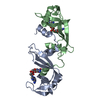
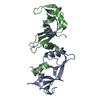
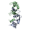
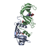
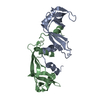
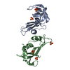
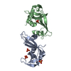
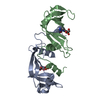
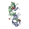
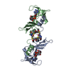
| Title | BOVINE SEMINAL RIBONUCLEASE STRUCTURE AT 1.9 ANGSTROMS RESOLUTION | ||||||
 Components Components | BOVINE SEMINAL RIBONUCLEASE | ||||||
 Keywords Keywords | HYDROLASE(PHOSPHORIC DIESTER / RNA) | ||||||
| Function / homology |  Function and homology information Function and homology informationpancreatic ribonuclease / ribonuclease A activity / RNA nuclease activity / nucleic acid binding / defense response to Gram-positive bacterium / hydrolase activity / extracellular region / identical protein binding Similarity search - Function | ||||||
| Biological species |  | ||||||
| Method |  X-RAY DIFFRACTION / Resolution: 1.9 Å X-RAY DIFFRACTION / Resolution: 1.9 Å | ||||||
 Authors Authors | Mazzarella, L. | ||||||
 Citation Citation | Journal: Acta Crystallogr.,Sect.D / Year: 1993 Title: Bovine seminal ribonuclease: structure at 1.9 A resolution. Authors: Mazzarella, L. / Capasso, S. / Demasi, D. / Di Lorenzo, G. / Mattia, C.A. / Zagari, A. #1: Journal: Gazz.Chim.Ital. / Year: 1987Title: Composite Active Sites in Bovine Seminal Ribonuclease Authors: Mazzarella, L. / Mattia, C.A. / Capasso, S. / Di Lorenzo, G. #2: Journal: Biopolymers / Year: 1983Title: Refinement of the Structure of Bovine Seminal Ribonuclease Authors: Capasso, S. / Giordano, F. / Mattia, C.A. / Mazzarella, L. / Zagari, A. #3: Journal: Gazz.Chim.Ital. / Year: 1979Title: Low-Resolution Structure of Bovine Seminal Ribonuclease: A Covalent Dimeric Protein Authors: Capasso, S. / Giordano, F. / Mattia, C.A. / Mazzarella, L. / Zagari, A. | ||||||
| History |
| ||||||
| Remark 700 | SHEET THE TWO BETA SHEETS OF EACH CHAIN OF THIS STRUCTURE ARE VERY CLOSE TO THOSE OF RNASE A (5RSA, ...SHEET THE TWO BETA SHEETS OF EACH CHAIN OF THIS STRUCTURE ARE VERY CLOSE TO THOSE OF RNASE A (5RSA, REMARK 5), AND A SIMILAR NOTATION HAS BEEN USED, MODIFIED ONLY TO TAKE INTO ACCOUNT THE PRESENCE OF TWO CHAINS. THEREFORE, DUE TO A BULGE OF RESIDUES 88 - 89, THE FIRST SHEET OF CHAIN A (B), COMPOSED OF 3 STRANDS, CONSISTS OF TWO PARTS DENOTED *S1A* AND *S2A* (*S1B* AND *S2B*), HAVING STRAND 1 AND 3 IDENTICAL. FOR CHAIN A (B), ALSO THE SECOND SHEET, FORMED BY 4 STRANDS, IS COMPOSED BY TWO PARTS DENOTED *S3A* AND *S4A* (*S3B* AND *S4B*), HAVING STRANDS 1, 2 AND 3 IDENTICAL. |
- Structure visualization
| Structure viewer | Molecule: MolmilJmol/JSmol |
|---|
- Downloads & links
-Download
| PDBx/mmCIF format | 1bsr.cif.gz | 62.7 KB | Display | PDBx/mmCIF format |
|---|---|---|---|---|
| PDB format | pdb1bsr.ent.gz | 46.9 KB | Display | PDB format |
| PDBx/mmJSON format | 1bsr.json.gz | Tree view | PDBx/mmJSON format | |
| Others |  Other downloads Other downloads |
-Validation report
| Arichive directory | https://data.pdbj.org/pub/pdb/validation_reports/bs/1bsrftp://data.pdbj.org/pub/pdb/validation_reports/bs/1bsr | HTTPS FTP |
|---|
-Related structure data
| Similar structure data |
|---|
-Links
 PDBj
PDBj

- Assembly
| Deposited unit | 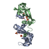
| ||||||||
|---|---|---|---|---|---|---|---|---|---|
| 1 |
| ||||||||
| 2 | 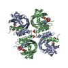
| ||||||||
| Unit cell |
| ||||||||
| Atom site foot note | 1: CIS PROLINE - PRO A 93 / 2: CIS PROLINE - PRO A 114 / 3: CIS PROLINE - PRO B 93 / 4: CIS PROLINE - PRO B 114 | ||||||||
| Components on special symmetry positions |
|
-Components
| #1: Protein | Mass: 13632.640 Da / Num. of mol.: 2 Source method: isolated from a genetically manipulated source Source: (gene. exp.) #2: Chemical | ChemComp-SO4 /   Mass: 96.063 Da / Num. of mol.: 7 / Source method: obtained synthetically / Formula: SO4 Mass: 96.063 Da / Num. of mol.: 7 / Source method: obtained synthetically / Formula: SO4#3: Water | ChemComp-HOH / |  Mass: 18.015 Da / Num. of mol.: 113 / Source method: isolated from a natural source / Formula: H2O Mass: 18.015 Da / Num. of mol.: 113 / Source method: isolated from a natural source / Formula: H2OCompound details | FEATURES OF THE STRUCTURE: RESIDUES 1 - 15 OF ONE CHAIN FOLD AGAINST RESIDUES 23 - 124 OF THE OTHER ...FEATURES OF THE STRUCTURE: RESIDUES 1 - 15 OF ONE CHAIN FOLD AGAINST RESIDUES 23 - 124 OF THE OTHER CHAIN. THEREFORE, IN THE DIMER, THE TWO CHAINS PRESENT THEIR N-TERMINI INTERCHANG | Has protein modification | Y | Nonpolymer details | A TOTAL OF 113 WATER MOLECULES, AS WELL AS 7 SULFATE ANIONS, WERE INCLUDED AND REFINED AS PART OF ...A TOTAL OF 113 WATER MOLECULES, AS WELL AS 7 SULFATE ANIONS, WERE INCLUDED AND REFINED AS PART OF THE STRUCTURE. | |
|---|
-Experimental details
-Experiment
| Experiment | Method: X-RAY DIFFRACTION |
|---|
- Sample preparation
| Crystal | Density Matthews: 2.4 Å3/Da / Density % sol: 48.72 % | |||||||||||||||||||||||||
|---|---|---|---|---|---|---|---|---|---|---|---|---|---|---|---|---|---|---|---|---|---|---|---|---|---|---|
| Crystal grow | *PLUS pH: 5.1 / Method: batch method | |||||||||||||||||||||||||
| Components of the solutions | *PLUS
|
-Data collection
| Radiation | Scattering type: x-ray |
|---|---|
| Radiation wavelength | Relative weight: 1 |
| Reflection | *PLUS Highest resolution: 1.9 Å / Lowest resolution: 9999 Å / Num. obs: 17217 / % possible obs: 90 % / Observed criterion σ(I): 3 / Num. measured all: 59869 / Rmerge(I) obs: 0.067 / Biso Wilson estimate: 9.2 Å2 |
- Processing
| Software |
| ||||||||||||||||||||||||||||||||||||||||||||||||||||||||||||
|---|---|---|---|---|---|---|---|---|---|---|---|---|---|---|---|---|---|---|---|---|---|---|---|---|---|---|---|---|---|---|---|---|---|---|---|---|---|---|---|---|---|---|---|---|---|---|---|---|---|---|---|---|---|---|---|---|---|---|---|---|---|
| Refinement | Rfactor Rwork: 0.177 / Rfactor obs: 0.177 / Highest resolution: 1.9 Å Details: A SECOND POSITION HAS BEEN LOCATED AND REFINED FOR THE SIDE CHAINS OF HIS A 119 AND ARG B 71. IN BOTH CHAINS, GLN 60 HAS A CONFORMATION WHICH FALLS OUTSIDE THE ALLOWED REGIONS OF THE ...Details: A SECOND POSITION HAS BEEN LOCATED AND REFINED FOR THE SIDE CHAINS OF HIS A 119 AND ARG B 71. IN BOTH CHAINS, GLN 60 HAS A CONFORMATION WHICH FALLS OUTSIDE THE ALLOWED REGIONS OF THE RAMACHANDRAN MAP, AS OBSERVED IN RNASE A. | ||||||||||||||||||||||||||||||||||||||||||||||||||||||||||||
| Refinement step | Cycle: LAST / Highest resolution: 1.9 Å
| ||||||||||||||||||||||||||||||||||||||||||||||||||||||||||||
| Refine LS restraints |
| ||||||||||||||||||||||||||||||||||||||||||||||||||||||||||||
| Refinement | *PLUS Highest resolution: 1.9 Å / Lowest resolution: 6 Å / Num. reflection obs: 16492 / σ(I): 3 / Rfactor obs: 0.177 | ||||||||||||||||||||||||||||||||||||||||||||||||||||||||||||
| Solvent computation | *PLUS | ||||||||||||||||||||||||||||||||||||||||||||||||||||||||||||
| Displacement parameters | *PLUS | ||||||||||||||||||||||||||||||||||||||||||||||||||||||||||||
| Refine LS restraints | *PLUS Type: x_angle_d / Dev ideal: 3.7 |
