 Movie
Movie Controller

[English] 日本語
 Yorodumi
- PDB-3f8g: The X-ray structure of a dimeric variant of human pancreatic ribo... -
+ Open data

- Basic information
| Entry | Database: PDB / ID: 3f8g | ||||||
|---|---|---|---|---|---|---|---|
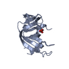
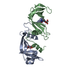
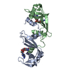
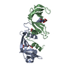
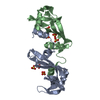
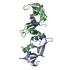
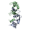
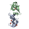
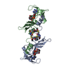
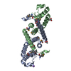
| Title | The X-ray structure of a dimeric variant of human pancreatic ribonuclease with high cytotoxic and antitumor activities | ||||||
 Components Components | Ribonuclease pancreatic | ||||||
 Keywords Keywords | HYDROLASE / antitumor agent / ribonuclease / 3d domain swapping / unswapped dimer / dimers / Endonuclease / Glycoprotein / Nuclease / Secreted | ||||||
| Function / homology |  Function and homology information Function and homology informationDevelopmental Lineage of Pancreatic Acinar Cells / pancreatic ribonuclease / ribonuclease A activity / RNA nuclease activity / Late endosomal microautophagy / Chaperone Mediated Autophagy / nucleic acid binding / defense response to Gram-positive bacterium / hydrolase activity / extracellular exosome Similarity search - Function | ||||||
| Biological species |  Homo sapiens (human) Homo sapiens (human) | ||||||
| Method |  X-RAY DIFFRACTION / SYNCHROTRON / MOLECULAR REPLACEMENT / Resolution: 2.6 Å X-RAY DIFFRACTION / SYNCHROTRON / MOLECULAR REPLACEMENT / Resolution: 2.6 Å | ||||||
 Authors Authors | Merlino, A. / Avella, G. / Mazzarella, L. / Sica, F. | ||||||
 Citation Citation | Journal: Protein Sci. / Year: 2009 Title: Structural features for the mechanism of antitumor action of a dimeric human pancreatic ribonuclease variant. Authors: Merlino, A. / Avella, G. / Di Gaetano, S. / Arciello, A. / Piccoli, R. / Mazzarella, L. / Sica, F. #1: Journal: J.Biol.Chem. / Year: 2004Title: Structure and stability of the non-covalent swapped dimer of bovine seminal ribonuclease: an enzyme tailored to evade ribonuclease protein inhibitor. Authors: Sica, F. / Di Fiore, A. / Merlino, A. / Mazzarella, L. #2: Journal: J.Mol.Biol. / Year: 2008Title: The buried diversity of bovine seminal ribonuclease: shape and cytotoxicity of the swapped non-covalent form of the enzyme. Authors: Merlino, A. / Ercole, C. / Picone, D. / Pizzo, E. / Mazzarella, L. / Sica, F. #3: Journal: Proc.Natl.Acad.Sci.USA / Year: 1999 Title: A dimeric mutant of human pancreatic ribonuclease with selective cytotoxicity toward malignant cells. Authors: Piccoli, R. / Di Gaetano, S. / De Lorenzo, C. / Grauso, M. / Monaco, C. / Spalletti-Cernia, D. / Laccetti, P. / Cinatl, J. / Matousek, J. / D'Alessio, G. #4: Journal: Biochem.J. / Year: 2001 Title: Second generation antitumour human RNase: significance of its structural and functional features for the mechanism of antitumour action. Authors: Di Gaetano, S. / D'alessio, G. / Piccoli, R. | ||||||
| History |
|
- Structure visualization
| Structure viewer | Molecule: MolmilJmol/JSmol |
|---|
- Downloads & links
-Download
| PDBx/mmCIF format | 3f8g.cif.gz | 65.2 KB | Display | PDBx/mmCIF format |
|---|---|---|---|---|
| PDB format | pdb3f8g.ent.gz | 48.6 KB | Display | PDB format |
| PDBx/mmJSON format | 3f8g.json.gz | Tree view | PDBx/mmJSON format | |
| Others |  Other downloads Other downloads |
-Validation report
| Arichive directory | https://data.pdbj.org/pub/pdb/validation_reports/f8/3f8gftp://data.pdbj.org/pub/pdb/validation_reports/f8/3f8g | HTTPS FTP |
|---|
-Related structure data
| Related structure data | 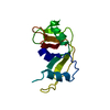 1e21S S: Starting model for refinement |
|---|---|
| Similar structure data |


-Links
 PDBj
PDBj

- Assembly
| Deposited unit | 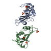
| ||||||||
|---|---|---|---|---|---|---|---|---|---|
| 1 |
| ||||||||
| Unit cell |
|
-Components
| #1: Protein | Mass: 14114.071 Da / Num. of mol.: 2 / Mutation: Q28L, R31C, R32C, N34K, E111G Source method: isolated from a genetically manipulated source Source: (gene. exp.) Homo sapiens (human) / Gene: RNASE1, RIB1, RNS1 / Production host:  #2: Chemical | ChemComp-SO4 /   Mass: 96.063 Da / Num. of mol.: 5 / Source method: obtained synthetically / Formula: SO4 Mass: 96.063 Da / Num. of mol.: 5 / Source method: obtained synthetically / Formula: SO4#3: Water | ChemComp-HOH / |  Mass: 18.015 Da / Num. of mol.: 162 / Source method: isolated from a natural source / Formula: H2O Mass: 18.015 Da / Num. of mol.: 162 / Source method: isolated from a natural source / Formula: H2OHas protein modification | Y | |
|---|
-Experimental details
-Experiment
| Experiment | Method: X-RAY DIFFRACTION / Number of used crystals: 1 |
|---|
- Sample preparation
| Crystal | Density Matthews: 2.56 Å3/Da / Density % sol: 52.04 % |
|---|---|
| Crystal grow | Temperature: 277 K / Method: vapor diffusion, sitting drop Details: A protein solution of 10mg/ml in 10mM Tris acteate ph 7.0, 300mM NaCl was equilibrated against a solution containing 27% w/v PEG, 0.2 M AS, and 100mM cacodylate buffer ph 6.5, VAPOR ...Details: A protein solution of 10mg/ml in 10mM Tris acteate ph 7.0, 300mM NaCl was equilibrated against a solution containing 27% w/v PEG, 0.2 M AS, and 100mM cacodylate buffer ph 6.5, VAPOR DIFFUSION, SITTING DROP, temperature 277K |
-Data collection
| Diffraction | Mean temperature: 100 K |
|---|---|
| Diffraction source | Source: SYNCHROTRON / Site: ELETTRA  / Beamline: 5.2R / Wavelength: 1 Å / Beamline: 5.2R / Wavelength: 1 Å |
| Detector | Type: MAR CCD 165 mm / Detector: CCD / Date: Oct 14, 2005 / Details: mirrors |
| Radiation | Monochromator: GRAPHITE / Protocol: SINGLE WAVELENGTH / Monochromatic (M) / Laue (L): M / Scattering type: x-ray |
| Radiation wavelength | Wavelength: 1 Å / Relative weight: 1 |
| Reflection | Resolution: 2.6→40 Å / Num. obs: 9128 / % possible obs: 98.7 % / Observed criterion σ(F): 0 / Observed criterion σ(I): 0 / Rmerge(I) obs: 0.09 / Net I/σ(I): 20 |
- Processing
| Software |
| |||||||||||||||||||||||||
|---|---|---|---|---|---|---|---|---|---|---|---|---|---|---|---|---|---|---|---|---|---|---|---|---|---|---|
| Refinement | Method to determine structure: MOLECULAR REPLACEMENT Starting model: PDB entry 1E21, the crystal structure of des(1-7)RNase Resolution: 2.6→40 Å / Isotropic thermal model: Isotropic / Cross valid method: THROUGHOUT / σ(F): 2 / Stereochemistry target values: Engh & Huber
| |||||||||||||||||||||||||
| Refinement step | Cycle: LAST / Resolution: 2.6→40 Å
|
