 Movie
Movie Controller


+ Open data

- Basic information
| Entry | Database: PDB / ID: 1.0E+21 | ||||||
|---|---|---|---|---|---|---|---|
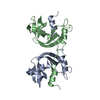
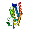
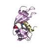
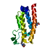
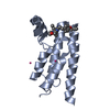
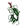
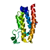
| Title | Ribonuclease 1 des1-7 Crystal Structure at 1.9A | ||||||
 Components Components | RIBONUCLEASE 1 | ||||||
 Keywords Keywords | HYDROLASE / HUMAN PANCREATIC RIBONUCLEASE | ||||||
| Function / homology |  Function and homology information Function and homology informationDevelopmental Lineage of Pancreatic Acinar Cells / pancreatic ribonuclease / ribonuclease A activity / RNA nuclease activity / Late endosomal microautophagy / Chaperone Mediated Autophagy / nucleic acid binding / defense response to Gram-positive bacterium / hydrolase activity / extracellular exosome Similarity search - Function | ||||||
| Biological species |  HOMO SAPIENS (human) HOMO SAPIENS (human) | ||||||
| Method |  X-RAY DIFFRACTION / SYNCHROTRON / MOLECULAR REPLACEMENT / Resolution: 1.9 Å X-RAY DIFFRACTION / SYNCHROTRON / MOLECULAR REPLACEMENT / Resolution: 1.9 Å | ||||||
 Authors Authors | Pous, J. / Mallorqui-Fernandez, G. / Peracaula, R. / Terzyan, S.S. / Futami, J. / Tada, H. / Yamada, H. / Seno, M. / De Llorens, R. / Gomis-Ruth, F.X. / Coll, M. | ||||||
 Citation Citation | Journal: Acta Crystallogr.,Sect.D / Year: 2001 Title: Three-Dimensional Crystal Structure of Human Rnase 1Dn7 at 1.9A Resolution Authors: Pous, J. / Mallorqui-Fernandez, G. / Peracaula, R. / Terzyan, S.S. / Futami, J. / Tada, H. / Yamada, H. / Seno, M. / De Llorens, R. / Gomis-Ruth, F.X. / Coll, M. | ||||||
| History |
|
- Structure visualization
| Structure viewer | Molecule: MolmilJmol/JSmol |
|---|
- Downloads & links
-Download
| PDBx/mmCIF format | 1e21.cif.gz | 39.5 KB | Display | PDBx/mmCIF format |
|---|---|---|---|---|
| PDB format | pdb1e21.ent.gz | 27.1 KB | Display | PDB format |
| PDBx/mmJSON format | 1e21.json.gz | Tree view | PDBx/mmJSON format | |
| Others |  Other downloads Other downloads |
-Validation report
| Arichive directory | https://data.pdbj.org/pub/pdb/validation_reports/e2/1e21ftp://data.pdbj.org/pub/pdb/validation_reports/e2/1e21 | HTTPS FTP |
|---|
-Related structure data
| Related structure data |  7rsaS S: Starting model for refinement |
|---|---|
| Similar structure data |


-Links
 PDBj
PDBj

- Assembly
| Deposited unit | 
| ||||||||
|---|---|---|---|---|---|---|---|---|---|
| 1 |
| ||||||||
| Unit cell |
|
-Components
| #1: Protein | Mass: 14539.300 Da / Num. of mol.: 1 / Mutation: YES Source method: isolated from a genetically manipulated source Source: (gene. exp.) HOMO SAPIENS (human) / Organ: PANCREAS / Plasmid: PET3A / Production host:  | ||
|---|---|---|---|
| #2: Water | ChemComp-HOH /  Mass: 18.015 Da / Num. of mol.: 137 / Source method: isolated from a natural source / Formula: H2O Mass: 18.015 Da / Num. of mol.: 137 / Source method: isolated from a natural source / Formula: H2O | ||
| Compound details | CHAIN A AMINO-TERMINALLY| Has protein modification | Y | |
-Experimental details
-Experiment
| Experiment | Method: X-RAY DIFFRACTION / Number of used crystals: 1 |
|---|
- Sample preparation
| Crystal | Density Matthews: 1.9 Å3/Da / Density % sol: 28 % | ||||||||||||||||||||
|---|---|---|---|---|---|---|---|---|---|---|---|---|---|---|---|---|---|---|---|---|---|
| Crystal grow | pH: 8.5 / Details: 7% PEG 8K, 0.1M TRIS-HCL PH 8.5 | ||||||||||||||||||||
| Crystal grow | *PLUS Method: vapor diffusion, hanging drop | ||||||||||||||||||||
| Components of the solutions | *PLUS
|
-Data collection
| Diffraction | Mean temperature: 100 K |
|---|---|
| Diffraction source | Source: SYNCHROTRON / Site: ESRF  / Beamline: ID14-2 / Wavelength: 0.933 / Beamline: ID14-2 / Wavelength: 0.933 |
| Detector | Type: ADSC CCD / Detector: CCD / Date: Feb 15, 2000 Details: SAGITALLY FOCUSING GE(220 MULTILAYER TOROIDAL MIRROR |
| Radiation | Monochromator: DIAMOND (111), GE(220) / Protocol: SINGLE WAVELENGTH / Monochromatic (M) / Laue (L): M / Scattering type: x-ray |
| Radiation wavelength | Wavelength: 0.933 Å / Relative weight: 1 |
| Reflection | Resolution: 1.9→20 Å / Num. obs: 8618 / % possible obs: 92.9 % / Observed criterion σ(I): 2 / Redundancy: 3.2 % / Biso Wilson estimate: 18.8 Å2 / Rmerge(I) obs: 0.135 / Rsym value: 0.113 / Net I/σ(I): 4.4 |
| Reflection shell | Resolution: 1.9→2 Å / Redundancy: 3.1 % / Rmerge(I) obs: 0.426 / Mean I/σ(I) obs: 2 / Rsym value: 0.364 / % possible all: 87.2 |
| Reflection | *PLUS Num. measured all: 27390 |
| Reflection shell | *PLUS % possible obs: 87.2 % / Rmerge(I) obs: 0.364 |
- Processing
| Software |
| ||||||||||||||||||||||||||||||||||||||||||||||||||||||||||||
|---|---|---|---|---|---|---|---|---|---|---|---|---|---|---|---|---|---|---|---|---|---|---|---|---|---|---|---|---|---|---|---|---|---|---|---|---|---|---|---|---|---|---|---|---|---|---|---|---|---|---|---|---|---|---|---|---|---|---|---|---|---|
| Refinement | Method to determine structure: MOLECULAR REPLACEMENT Starting model: PDB ENTRY 7RSA Resolution: 1.9→20 Å / Data cutoff high absF: 10000 / Cross valid method: THROUGHOUT / σ(F): 0 / Details: RESIDUES 126, 127 AND 128 NOT SEEN IN DENSITY MAPS
| ||||||||||||||||||||||||||||||||||||||||||||||||||||||||||||
| Solvent computation | Bsol: 50.93 Å2 / ksol: 0.34 e/Å3 | ||||||||||||||||||||||||||||||||||||||||||||||||||||||||||||
| Displacement parameters | Biso mean: 23.496 Å2 | ||||||||||||||||||||||||||||||||||||||||||||||||||||||||||||
| Refinement step | Cycle: LAST / Resolution: 1.9→20 Å
| ||||||||||||||||||||||||||||||||||||||||||||||||||||||||||||
| Refine LS restraints |
| ||||||||||||||||||||||||||||||||||||||||||||||||||||||||||||
| LS refinement shell | Resolution: 1.9→1.99 Å / Total num. of bins used: 8
| ||||||||||||||||||||||||||||||||||||||||||||||||||||||||||||
| Software | *PLUS Name: CNS / Version: 1 / Classification: refinement | ||||||||||||||||||||||||||||||||||||||||||||||||||||||||||||
| LS refinement shell | *PLUS Rfactor Rfree: 0.298 |
