 Movie
Movie Controller


+ Open data

- Basic information
| Entry | Database: PDB / ID: 1h8x | ||||||
|---|---|---|---|---|---|---|---|
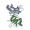
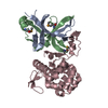
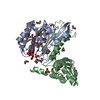
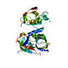
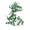
| Title | Domain-swapped Dimer of a Human Pancreatic Ribonuclease Variant | ||||||
 Components Components | RIBONUCLEASE 1 | ||||||
 Keywords Keywords | HYDROLASE / RIBONUCLEASE / RNASE / HUMAN PANCREATIC RIBONUCLEASE / DOMAIN- SWAPPED DIMER | ||||||
| Function / homology |  Function and homology information Function and homology informationDevelopmental Lineage of Pancreatic Acinar Cells / pancreatic ribonuclease / ribonuclease A activity / RNA nuclease activity / Late endosomal microautophagy / Chaperone Mediated Autophagy / nucleic acid binding / defense response to Gram-positive bacterium / hydrolase activity / extracellular exosome Similarity search - Function | ||||||
| Biological species |  HOMO SAPIENS (human) HOMO SAPIENS (human) | ||||||
| Method |  X-RAY DIFFRACTION / MOLECULAR REPLACEMENT / Resolution: 2 Å X-RAY DIFFRACTION / MOLECULAR REPLACEMENT / Resolution: 2 Å | ||||||
 Authors Authors | Canals, A. / Pous, J. / Guasch, A. / Benito, A. / Ribo, M. / Vilanova, M. / Coll, M. | ||||||
 Citation Citation | Journal: Structure / Year: 2001 Title: The Structure of an Engineered Domain-Swapped Ribonuclease Dimer and its Implications for the Evolution of Proteins Toward Oligomerization Authors: Canals, A. / Pous, J. / Guasch, A. / Benito, A. / Ribo, M. / Vilanova, M. / Coll, M. | ||||||
| History |
|
- Structure visualization
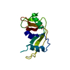
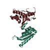
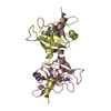
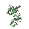
| Structure viewer | Molecule: MolmilJmol/JSmol |
|---|
- Downloads & links
-Download
| PDBx/mmCIF format | 1h8x.cif.gz | 70.1 KB | Display | PDBx/mmCIF format |
|---|---|---|---|---|
| PDB format | pdb1h8x.ent.gz | 52.4 KB | Display | PDB format |
| PDBx/mmJSON format | 1h8x.json.gz | Tree view | PDBx/mmJSON format | |
| Others |  Other downloads Other downloads |
-Validation report
| Arichive directory | https://data.pdbj.org/pub/pdb/validation_reports/h8/1h8xftp://data.pdbj.org/pub/pdb/validation_reports/h8/1h8x | HTTPS FTP |
|---|
-Related structure data
| Related structure data |  1dzaS S: Starting model for refinement |
|---|---|
| Similar structure data |


-Links
 PDBj
PDBj

- Assembly
| Deposited unit | 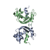
| ||||||||
|---|---|---|---|---|---|---|---|---|---|
| 1 |
| ||||||||
| Unit cell |
|
-Components
| #1: Protein | Mass: 14454.169 Da / Num. of mol.: 2 / Fragment: RNASE 1, HP-RNASE / Mutation: YES Source method: isolated from a genetically manipulated source Details: RESIDIE 100 IS A FORMILMETHIONINE (FME) / Source: (gene. exp.) HOMO SAPIENS (human) / Description: SYNTHETIC GENE / Gene: PM8 / Organ: PANCREAS / Plasmid: PM8 / Gene (production host): PM8 / Production host:  #2: Water | ChemComp-HOH / |  Mass: 18.015 Da / Num. of mol.: 340 / Source method: isolated from a natural source / Formula: H2O Mass: 18.015 Da / Num. of mol.: 340 / Source method: isolated from a natural source / Formula: H2OCompound details | CHAIN A, B ENGINEERED | Has protein modification | Y | |
|---|
-Experimental details
-Experiment
| Experiment | Method: X-RAY DIFFRACTION / Number of used crystals: 1 |
|---|
- Sample preparation
| Crystal | Density Matthews: 2.13 Å3/Da / Density % sol: 30 % | ||||||||||||||||||||||||
|---|---|---|---|---|---|---|---|---|---|---|---|---|---|---|---|---|---|---|---|---|---|---|---|---|---|
| Crystal grow | pH: 9 / Details: pH 9.00 | ||||||||||||||||||||||||
| Crystal grow | *PLUS Method: vapor diffusion, hanging drop | ||||||||||||||||||||||||
| Components of the solutions | *PLUS
|
-Data collection
| Diffraction | Mean temperature: 110 K |
|---|---|
| Diffraction source | Source: ROTATING ANODE / Type: RIGAKU RU200 / Wavelength: 1.5418 |
| Detector | Type: MARRESEARCH / Detector: IMAGE PLATE / Date: Feb 15, 1999 |
| Radiation | Protocol: SINGLE WAVELENGTH / Monochromatic (M) / Laue (L): M / Scattering type: x-ray |
| Radiation wavelength | Wavelength: 1.5418 Å / Relative weight: 1 |
| Reflection | Resolution: 1.9→13 Å / Num. obs: 17315 / % possible obs: 99.4 % / Redundancy: 3.1 % / Biso Wilson estimate: 33.7 Å2 / Rmerge(I) obs: 0.076 / Net I/σ(I): 7.1 |
| Reflection shell | Resolution: 1.9→2 Å / Redundancy: 3.1 % / Rmerge(I) obs: 0.315 / Mean I/σ(I) obs: 2.4 / % possible all: 99.8 |
| Reflection | *PLUS Highest resolution: 2 Å / Lowest resolution: 13 Å / Num. obs: 16247 / Redundancy: 3.4 % / Num. measured all: 56729 |
| Reflection shell | *PLUS Highest resolution: 2 Å / Lowest resolution: 2.12 Å |
- Processing
| Software |
| ||||||||||||||||||||||||||||||||||||||||||||||||||||||||||||||||||||||||||||||||
|---|---|---|---|---|---|---|---|---|---|---|---|---|---|---|---|---|---|---|---|---|---|---|---|---|---|---|---|---|---|---|---|---|---|---|---|---|---|---|---|---|---|---|---|---|---|---|---|---|---|---|---|---|---|---|---|---|---|---|---|---|---|---|---|---|---|---|---|---|---|---|---|---|---|---|---|---|---|---|---|---|---|
| Refinement | Method to determine structure: MOLECULAR REPLACEMENT Starting model: PDB ENTRY 1DZA Resolution: 2→12 Å / Isotropic thermal model: RESTRAINED / Cross valid method: THROUGHOUT / σ(F): 0
| ||||||||||||||||||||||||||||||||||||||||||||||||||||||||||||||||||||||||||||||||
| Solvent computation | Solvent model: DENSITY MODIFICATION | ||||||||||||||||||||||||||||||||||||||||||||||||||||||||||||||||||||||||||||||||
| Displacement parameters | Biso mean: 29.3 Å2 | ||||||||||||||||||||||||||||||||||||||||||||||||||||||||||||||||||||||||||||||||
| Refine analyze |
| ||||||||||||||||||||||||||||||||||||||||||||||||||||||||||||||||||||||||||||||||
| Refinement step | Cycle: LAST / Resolution: 2→12 Å
| ||||||||||||||||||||||||||||||||||||||||||||||||||||||||||||||||||||||||||||||||
| Refine LS restraints |
| ||||||||||||||||||||||||||||||||||||||||||||||||||||||||||||||||||||||||||||||||
| LS refinement shell | Resolution: 2→2.07 Å / Total num. of bins used: 10
| ||||||||||||||||||||||||||||||||||||||||||||||||||||||||||||||||||||||||||||||||
| Xplor file |
| ||||||||||||||||||||||||||||||||||||||||||||||||||||||||||||||||||||||||||||||||
| Software | *PLUS Name: CNS / Version: 1 / Classification: refinement | ||||||||||||||||||||||||||||||||||||||||||||||||||||||||||||||||||||||||||||||||
| Refinement | *PLUS Highest resolution: 2 Å | ||||||||||||||||||||||||||||||||||||||||||||||||||||||||||||||||||||||||||||||||
| Solvent computation | *PLUS | ||||||||||||||||||||||||||||||||||||||||||||||||||||||||||||||||||||||||||||||||
| Displacement parameters | *PLUS | ||||||||||||||||||||||||||||||||||||||||||||||||||||||||||||||||||||||||||||||||
| Refine LS restraints | *PLUS
| ||||||||||||||||||||||||||||||||||||||||||||||||||||||||||||||||||||||||||||||||
| LS refinement shell | *PLUS Rfactor Rwork: 0.27 |
