 Movie
Movie Controller

[English] 日本語
 Yorodumi
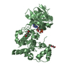
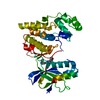
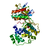
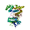
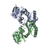
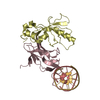
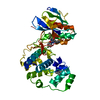
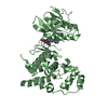
- PDB-4yxa: Complex of SpaO(SPOA1,2 SeMet) and OrgB(APAR)::T4lysozyme fusion ... -
+ Open data

- Basic information
| Entry | Database: PDB / ID: 4yxa | ||||||
|---|---|---|---|---|---|---|---|
| Title | Complex of SpaO(SPOA1,2 SeMet) and OrgB(APAR)::T4lysozyme fusion protein | ||||||
 Components Components |
| ||||||
 Keywords Keywords | PROTEIN TRANSPORT / Type III Secretion System | ||||||
| Function / homology |  Function and homology information Function and homology informationprotein secretion by the type III secretion system / bacterial-type flagellum-dependent swarming motility / positive chemotaxis / viral release from host cell by cytolysis / peptidoglycan catabolic process / cell wall macromolecule catabolic process / lysozyme / lysozyme activity / protein transport / host cell cytoplasm ...protein secretion by the type III secretion system / bacterial-type flagellum-dependent swarming motility / positive chemotaxis / viral release from host cell by cytolysis / peptidoglycan catabolic process / cell wall macromolecule catabolic process / lysozyme / lysozyme activity / protein transport / host cell cytoplasm / defense response to bacterium / cytoplasm Similarity search - Function | ||||||
| Biological species |  Salmonella typhimurium (bacteria) Salmonella typhimurium (bacteria) Enterobacteria phage T4 (virus) Enterobacteria phage T4 (virus) | ||||||
| Method |  X-RAY DIFFRACTION / SYNCHROTRON / MOLECULAR REPLACEMENT / Resolution: 2.35 Å X-RAY DIFFRACTION / SYNCHROTRON / MOLECULAR REPLACEMENT / Resolution: 2.35 Å | ||||||
 Authors Authors | Notti, R.Q. / Stebbins, C.E. | ||||||
 Citation Citation | Journal: Nat Commun / Year: 2015 Title: A common assembly module in injectisome and flagellar type III secretion sorting platforms. Authors: Notti, R.Q. / Bhattacharya, S. / Lilic, M. / Stebbins, C.E. | ||||||
| History |
|
- Structure visualization
| Structure viewer | Molecule: MolmilJmol/JSmol |
|---|
- Downloads & links
-Download
| PDBx/mmCIF format | 4yxa.cif.gz | 137.3 KB | Display | PDBx/mmCIF format |
|---|---|---|---|---|
| PDB format | pdb4yxa.ent.gz | 106.8 KB | Display | PDB format |
| PDBx/mmJSON format | 4yxa.json.gz | Tree view | PDBx/mmJSON format | |
| Others |  Other downloads Other downloads |
-Validation report
| Arichive directory | https://data.pdbj.org/pub/pdb/validation_reports/yx/4yxaftp://data.pdbj.org/pub/pdb/validation_reports/yx/4yxa | HTTPS FTP |
|---|
-Related structure data
| Related structure data | 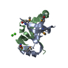 4yx1C 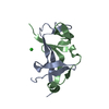 4yx5C 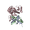 4yx7SC 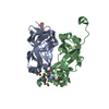 4yxbC 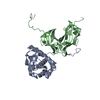 4yxcC S: Starting model for refinement C: citing same article ( |
|---|---|
| Similar structure data |


-Links
 PDBj
PDBj


- Assembly
| Deposited unit | 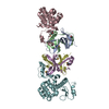
| ||||||||
|---|---|---|---|---|---|---|---|---|---|
| 1 | 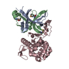
| ||||||||
| 2 | 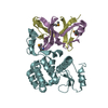
| ||||||||
| Unit cell |
|
-Components
| #1: Protein | Mass: 8246.572 Da / Num. of mol.: 2 / Fragment: UNP Residues 145-213 Source method: isolated from a genetically manipulated source Source: (gene. exp.) Salmonella typhimurium (strain LT2 / SGSC1412 / ATCC 700720) (bacteria)Strain: LT2 / SGSC1412 / ATCC 700720 / Gene: spaO, STM2891 / Production host: #2: Protein | Mass: 7851.493 Da / Num. of mol.: 2 / Fragment: UNP Residues 232-297 Source method: isolated from a genetically manipulated source Source: (gene. exp.) Salmonella typhimurium (bacteria) / Strain: LT2 / SGSC1412 / ATCC 700720 / Gene: spaO, STM2891 / Production host: #3: Protein | Mass: 22162.568 Da / Num. of mol.: 2 / Fragment: UNP Residues 1-30,UNP Residues 1-30 / Mutation: D20N, C54T, C97A,D20N, C54T, C97A Source method: isolated from a genetically manipulated source Source: (gene. exp.) Salmonella typhimurium (strain LT2 / SGSC1412 / ATCC 700720) (bacteria), (gene. exp.) Enterobacteria phage T4 (virus)Strain: LT2 / SGSC1412 / ATCC 700720 / Gene: orgB, STM2869 / Production host: #4: Water | ChemComp-HOH / |  Mass: 18.015 Da / Num. of mol.: 122 / Source method: isolated from a natural source / Formula: H2O Mass: 18.015 Da / Num. of mol.: 122 / Source method: isolated from a natural source / Formula: H2OHas protein modification | Y | |
|---|
-Experimental details
-Experiment
| Experiment | Method: X-RAY DIFFRACTION / Number of used crystals: 1 |
|---|
- Sample preparation
| Crystal | Density Matthews: 2.15 Å3/Da / Density % sol: 42.69 % |
|---|---|
| Crystal grow | Temperature: 293 K / Method: vapor diffusion, hanging drop Details: SpaO(145-213, SeMet) + SpaO (232-297, SeMet) + OrgB(1-30)::T4 lysozyme (native) was concentrated to 18mg/mL, supplemented with 50mM maltose, and crystallized with 25% PEG3350, 200mM ammonium ...Details: SpaO(145-213, SeMet) + SpaO (232-297, SeMet) + OrgB(1-30)::T4 lysozyme (native) was concentrated to 18mg/mL, supplemented with 50mM maltose, and crystallized with 25% PEG3350, 200mM ammonium formate, 100mM sodium acetate pH=5.0. Microseeding was employed to enhance crystal uniformity and diffraction. Briefly, crystals to be seeded were harvested in precipitant solution and vortexed in a microfuge tube with a small stir bar for ~60 seconds. The slurry of microseeds was serially dilluted (5-10-fold steps) in precipitant solution and 5 selected microseed-precipitant mixtures were mixed with fresh protein as in a normal hanging drop experiment. Crystals were cryoprotected in 25% PEG3350, 10% ethylene glycol, 200mM ammonium formate, 100mM sodium acetate pH=5.0, 50mM maltose. |
-Data collection
| Diffraction | Mean temperature: 100 K | |||||||||||||||||||||||||||
|---|---|---|---|---|---|---|---|---|---|---|---|---|---|---|---|---|---|---|---|---|---|---|---|---|---|---|---|---|
| Diffraction source | Source: SYNCHROTRON / Site: NSLS  / Beamline: X29A / Wavelength: 0.979 Å / Beamline: X29A / Wavelength: 0.979 Å | |||||||||||||||||||||||||||
| Detector | Type: ADSC QUANTUM 315r / Detector: CCD / Date: Mar 12, 2014 | |||||||||||||||||||||||||||
| Radiation | Protocol: SINGLE WAVELENGTH / Monochromatic (M) / Laue (L): M / Scattering type: x-ray | |||||||||||||||||||||||||||
| Radiation wavelength | Wavelength: 0.979 Å / Relative weight: 1 | |||||||||||||||||||||||||||
| Reflection twin | Operator: l,-k,h / Fraction: 0.28 | |||||||||||||||||||||||||||
| Reflection | Resolution: 2.35→47.61 Å / Num. obs: 25759 / % possible obs: 99 % / Redundancy: 6.6 % / CC1/2: 0.997 / Rmerge(I) obs: 0.088 / Rpim(I) all: 0.037 / Net I/σ(I): 12.8 / Num. measured all: 169948 / Scaling rejects: 92 | |||||||||||||||||||||||||||
| Reflection shell | Diffraction-ID: 1 / Rejects: _
|
- Processing
| Software |
| ||||||||||||||||||||||||||||||||||||||||||||||||||||||||||||||||||||||||||||||||||||
|---|---|---|---|---|---|---|---|---|---|---|---|---|---|---|---|---|---|---|---|---|---|---|---|---|---|---|---|---|---|---|---|---|---|---|---|---|---|---|---|---|---|---|---|---|---|---|---|---|---|---|---|---|---|---|---|---|---|---|---|---|---|---|---|---|---|---|---|---|---|---|---|---|---|---|---|---|---|---|---|---|---|---|---|---|---|
| Refinement | Method to determine structure: MOLECULAR REPLACEMENT Starting model: 4YX7 Resolution: 2.35→45.804 Å / Cross valid method: FREE R-VALUE / σ(F): 1.38 / Phase error: 35.43 / Stereochemistry target values: TWIN_LSQ_F
| ||||||||||||||||||||||||||||||||||||||||||||||||||||||||||||||||||||||||||||||||||||
| Solvent computation | Shrinkage radii: 0.9 Å / VDW probe radii: 1.11 Å / Solvent model: FLAT BULK SOLVENT MODEL | ||||||||||||||||||||||||||||||||||||||||||||||||||||||||||||||||||||||||||||||||||||
| Displacement parameters | Biso max: 132.27 Å2 / Biso mean: 46.8298 Å2 / Biso min: 19.12 Å2 | ||||||||||||||||||||||||||||||||||||||||||||||||||||||||||||||||||||||||||||||||||||
| Refinement step | Cycle: final / Resolution: 2.35→45.804 Å
| ||||||||||||||||||||||||||||||||||||||||||||||||||||||||||||||||||||||||||||||||||||
| Refine LS restraints |
| ||||||||||||||||||||||||||||||||||||||||||||||||||||||||||||||||||||||||||||||||||||
| LS refinement shell | Refine-ID: X-RAY DIFFRACTION / Total num. of bins used: 11
|
